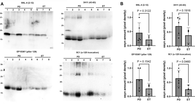
Fig. 5.
Western blot immunochemical detection of αSyn. (A) DBS samples purified using SDS were obtained from five PD samples (lanes 1–5) and three ET samples (lanes 6–8) and subject to immunoblotting comparison of αSyn species using a panel of four antibodies which are indicated. 5 µg of sample are loaded in all lanes for all samples. Antibody SNL-4 specific for residues 2–12 of αSyn demonstrates αSyn detection at ~ 17 kDa for all samples, antibody 3H11 specific for residues 43–63 of αSyn demonstrates αSyn along with additional heavier bands that may be oligomers. Antibody EP1536Y specific for pSer129 αSyn detects this epitope at ~ 17 kDa in all samples, and antibody 5C1 specific for truncated αSyn at 125 displays detection of ~ 14 kDa band preferentially in the PD samples. Band size in kDa is shown. (B) Densitometric analysis of band detection in PD (n = 5) and ET (n = 3) samples with each of the four antibodies utilized; comparison using T-test and associated P value displayed. 5C1 has significantly enhanced detection of truncated αSyn at 125 in PD samples compared with ET. Western blot images are displayed with increased brightness and contrast for ease of viewing, with originals in Supplementary information.

