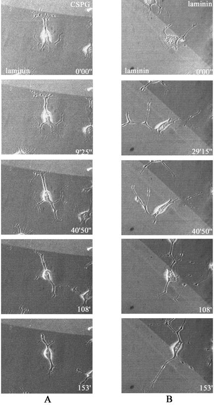
FIG. 10.
Time-lapse analysis of neurites stopping at the CSPG boundary. Outgrowing cell neurites stopping and retracting at a laminin-CSPG boundary (A) or freely crossing a laminin-laminin boundary, followed by crossing of the cell body itself (B). Neurite outgrowth was induced by serum starvation and treatment with 10 μM Y-27632. Details are given in Materials and Methods. The cells were subjected to time-lapse imaging at 45-s intervals for 2.5 h, with the first images taken 30 min after serum starvation/treatment. The phase-contrast images of cells were superimposed on fluorescent images of (A) CSPG-laminin or (B) laminin-laminin substrates, with TRITC included in the upper (A) or lower (B) substrates, using confocal microscopy.

