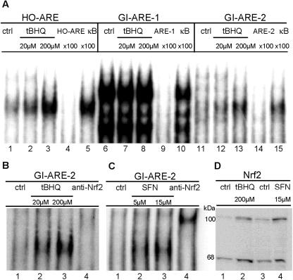
FIG. 2.
Nrf2 translocates to the nucleus and binds to GI-ARE-2 in response to tBHQ or SFN exposure. Nuclear extracts were prepared from HepG2 cells treated with tBHQ (20, 200 μM; 16 h) or SFN (5, 15 μM; 4 h). EMSAs were performed as described in Materials and Methods. (A) HO-ARE (lanes 1 to 5), GI-ARE-1 (lanes 6 to 10), and GI-ARE-2 (lanes 11 to 15) probes were incubated with nuclear extracts from stimulated cells as indicated. For control, a 100-fold molar excess of the respective unlabeled specific (lanes 4, 9, 14) or unspecific (κB; lanes 5, 10, 15) oligonucleotide was added during the binding procedure. (B and C) GI-ARE-2 was incubated with nuclear extracts of tBHQ-treated (B) or SFN-treated (C) cells. For supershift, nuclear extracts were incubated with anti-Nrf2 (lane 4). Results are representative of three independent experiments. (D) HepG2 cells were stimulated with tBHQ (200 μM, 16 h) or SFN (15 μM, 4 h). Nuclear extracts were analyzed for Nrf2 by Western blotting as described in Materials and Methods. Results are representative of three independent experiments.

