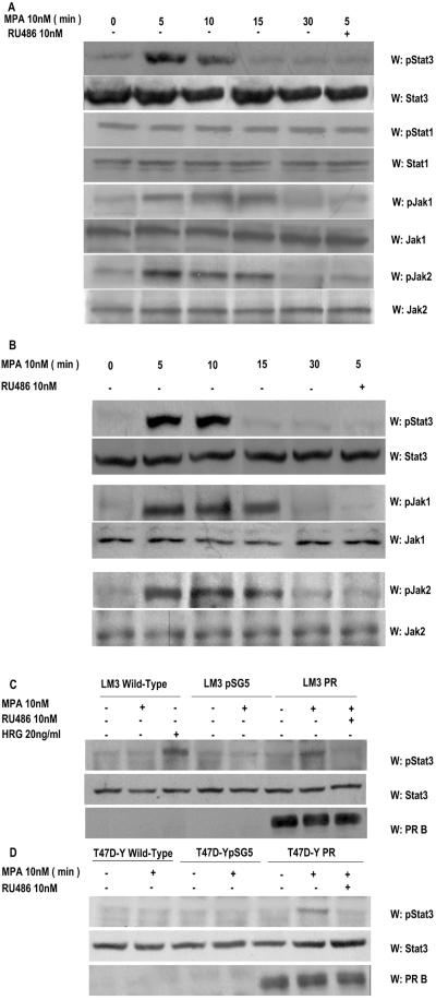
FIG. 2.
MPA induces tyrosine phosphorylation by Stat3, Jak1, and Jak2. Cultures of C4HD (A) and T47D (B) cells were treated with 10 nM MPA or MPA-10 nM RU486 for the indicated times. Fifty micrograms of protein from cell lysates was electrophoresed, and Western blot assays were performed with antiphosphotyrosine 705 Stat3, antiphosphotyrosine 701 Stat1, antiphosphotyrosine 1022/1023 Jak1, and antiphosphotyrosine 1007/1008 Jak2 antibodies. Membranes were then stripped and hybridized with anti-Stat3, -Stat1, -Jak1, and -Jak2 antibodies. This experiment was repeated six times for C4HD cells and three times for T47D cells with similar results. LM3 (C) or T47D-Y (D) cells were transfected with PRB or with the empty pSG5 plasmidor remained untreated. Cells were then stimulated for 5 min with MPA or pretreated with RU486 before MPA stimulation. LM3 cells were also treated with heregulin for 10 min. Fifty micrograms of protein from cell lysates was electrophoresed, and Western blot assays were performed with antiphosphotyrosine 705 Stat3 (upper parts of panels C and D). Membranes were then stripped and hybridized with anti-Stat3 (middle panes of parts C and D) and anti-PR (lower parts of panels C and D) antibodies. This experiment was repeated three times with similar results. W, Western blot assay.

