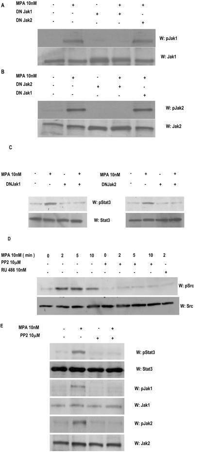
FIG. 3.
Jak1 and Jak2 are involved in MPA-induced Stat3 phosphorylation. C4HD cells were transiently transfected with 2 μg of DN Jak1 or DN Jak2 vector and then treated with MPA for 5 min or left untreated. Fifty micrograms of protein from cell lysates was electrophoresed, and Western blot assays were performed with antiphosphotyrosine Jak1 (A, upper part) or antiphosphotyrosine Jak2 (B, upper part) antibodies. Membranes were then stripped and hybridized with anti-Jak1 (A, lower part) and anti-Jak2 (B, lower part) antibodies. (C) Fifty micrograms of protein from cells transfected with the DN Jak1 (left part) or the DN Jak2 (right part) vector and subsequentlytreated with MPA for 5 min or left untreated was electrophoresed, and Western blot assays were performed with antiphosphotyrosine Stat3 (upper parts). Membranes were then stripped and hybridized with anti-Stat3 (lower parts) antibodies. (D) C4HD cells were treated with MPA for the indicated times or preincubated with the selective Src family kinase inhibitor PP2 or RU486 for 90 min and then treated with MPA. Fifty micrograms of protein from cell lysates was electrophoresed and immunoblotted with an antiphosphotyrosine c-Src antibody (upper part). Membrane was then stripped and hybridized with anti-c-Src antibody (lower part). (E) C4HD cells were preincubated with the selective Src family kinase inhibitor PP2 for 90 min and then treated with MPA for 5 min. Fifty micrograms of protein from cell lysates was electrophoresed and immunoblotted with antiphosphotyrosine Stat3, antiphosphotyrosine Jak1, and antiphosphotyrosine Jak2 antibodies. Membranes were then stripped and hybridized with anti-Stat3, anti-Jak1, and anti-Jak2 antibodies, respectively. These experiments were repeated three times with similar results. W, Western blot assay.

