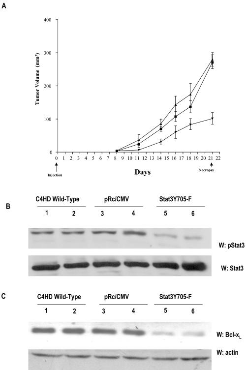
FIG. 9.
In vivo blockage of Stat3 expression. (A) C4HD cells growing in 10 nM MPA were transiently transfected with the DN Stat3Y705-F expression vector (▾) or the empty pRc/CMV vector (▪) or remained untreated (▴), as described in the legend to Fig. 8. After 48 h of transfection, 106 cells from each experimental group were inoculated s.c. into animals treated with a 40-mg MPA depot in the flank opposite the cell inoculum, and tumor width and length were measured three times a week in order to calculate volume as described in Materials and Methods. Each point represents the mean volume ± the standard error of nine independent tumors for each control group and of four tumors that developed in mice injected with Stat3Y705-F-transfected cells. (B) One hundred micrograms of protein from tumor lysates was electrophoresed and immunoblotted with an anti-phospho-Stat3 antibody (upper part). Shown are two representative samples of mice injected with C4HD wild-type cells (lanes 1 and 2), C4HD cells transfected with the empty pRc/CMV vector (lanes 3 and 4), and C4HD cells transfected with the DN Stat3Y705-F expression vector (lanes 5 and 6). Membrane was stripped and hybridized with an anti-Stat3 antibody (lower part). This is a representative experiment out of a total of three. Densitometric analysis of Stat3 phosphorylated bands from the four tumors that developed in mice injected with C4HD cells transfected with the DN Stat3Y705-F vector and from multiple C4HD tumors that developed in mice injected with either wild-type C4HD cells or with C4HD cells transfected with the empty pRc/CMV vector showed a significant decrease in Stat3 tyrosine phosphorylation in tumors from mice injected with cells transfected with the DN Stat3Y705-F vector, with respect to tumors growing in control animals (P < 0.001). W, Western blot assay. (C) One hundred micrograms of protein from tumor lysates was electrophoresed and immunoblotted with an anti-Bcl-xL antibody (upper part). Shown are two representative samples of mice injected with C4HD wild-type cells (lanes 1 and 2), with C4HD cells transfected with the empty pRc/CMV vector (lanes 3 and 4), andwith C4HD cells transfected with the DN Stat3Y705-F expression vector (lanes 5 and 6). Membrane was then stripped and hybridized with an antiactin antibody (lower part), as a control for the specificity of the DN Stat3Y705-F effect. Densitometric analysis of the Bcl-xL band from tumors that developed in mice injected with C4HD cells transfected with the DN Stat3Y705-F, expressed as a percentage of the control values (i.e., tumors growing in control groups), ranged between 25 and 40% for tumors growing in mice injected with DN Stat3Y705-F-transfected cells. There was significant inhibition of Bcl-xL expression in mice injected with DN Stat3Y705-F-transfected cells with respect to mice injected with empty pRc/CMV vector-transfected cells or with wild-type C4HD cells (P < 0.001).

