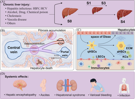
FIGURE 1.

The pathophysiology and molecular mechanism of liver cirrhosis. (A) Chronic liver injury caused by various etiologies leads to liver fibrosis. Liver biopsy is the gold standard for the diagnosis of liver fibrosis, and the histopathological staging mainly includes stage 0 (S0): no definite fibrosis; stage 1 (S1): mild fibrosis; stage 2 (S2): moderate fibrosis; stage 3 (S3): advanced liver fibrosis; and stage 4 (S4): liver cirrhosis. (B) Liver cirrhosis leads to intrahepatic resistance, including hepatocyte death and replacement by fibrotic tissue, structural changes in hepatic sinuses (e.g., regenerative nodules) leading to the restriction of sinusoidal flow, and functional contraction of intrahepatic blood vessels due to the reduced production of vasodilators (e.g., NO). These features disrupt the normal metabolic functions of the liver, resulting in hepatic dysfunction, portal hypertension, and other systemic effects. (C) Long‐term inflammation leads to an imbalance in the liver microenvironment, which is associated with cell heterogeneity, interactions with various cell types, and microenvironmental factors in liver tissue and stimulates the occurrence of liver cirrhosis. (D) The systemic effects of liver cirrhosis. This figure was generated using FigDraw (https://www.figdraw.com).
