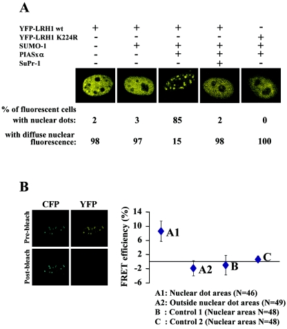
FIG. 3.
SUMO-modified LRH-1 is localized in discrete nuclear dots. (A) HeLa cells were transfected with the indicated expression vectors and observed by confocal microscopy. Representative images of cells from several experiments are shown. (B) HeLa cells were transfected with pECFP-SUMO-1, pEYFP-LRH-1, and pCMV-Flag-PIASxα. Sequential CFP and YFP fluorescence images were recorded before and after 1 min of photobleaching of YFP fluorescence by 514-nm laser line. FRET efficiency was expressed as the percent increase of prebleach CFP fluorescence after YFP photobleaching in the nuclear dot areas (A1) and similar-sized areas outside the nuclear dots (A2). In the control experiment B, the cells were cotransfected with pECFP-SUMO-1, pEYFP-LRH-1 K224R, and pCMV-Flag-PIASxα, whereas in the control experiment C, the cells were cotransfected with pECFP-SUMO-1 and pEYFP-LRH-1 without the PIASxα expression vector. The differences in fluorescence intensity in similar-sized nuclear areas were calculated as described above. The graph shows mean values and standard errors from the indicated number (N) of measurements, which were obtained from six to eight different cells.

