FIG. 5.
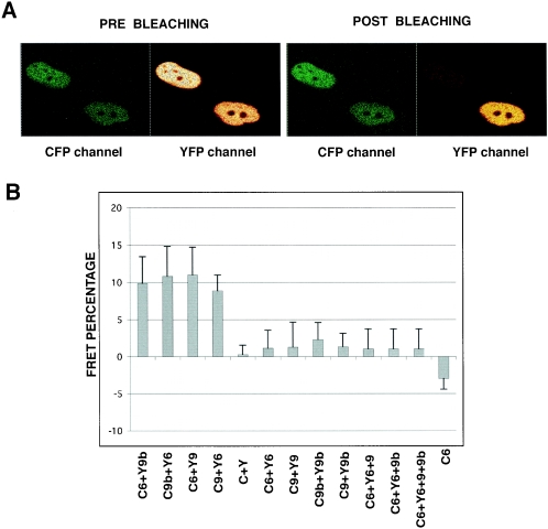
TAF9b interacts with TAF6, similarly to TAF9, in vivo. Acceptor photobleaching of cells transfected with the indicated combinations of vectors expressing YFP (Y) and CFP (C) fusion proteins with TAF6 (6), TAF9 (9), and TAF9b (9b). (A) Representative CFP-TAF6 (donor) channel and YFP-TAF9b (acceptor) channel images before and after bleaching are shown. Note the dramatic drop in YFP fluorescence and the increase in CFP fluorescence in the cell after bleaching by the 514-nm laser line (the cell in the upper left corner of the panels). The cell in the lower right corner of the panels was not bleached and serves as a control. (B) Mean values of FRET efficiencies following quantification of the increase of the CFP fluorescence intensity after bleaching in 60 cells from three different experiments. To calculate FRET efficiency in living cells, the following formula was used: EF = (Ipost − Ipre) × 100/Ipost. The mean values for the FRET efficiency of TAF6-TAF9 (C6+Y9 or Y6+C9) and TAF6-TAF9b (C6+Y9b or Y6+C9b) combinations (first four bars) are similar to each other and significantly different from the empty CFP-YFP vector combination (C+Y) or CFP-TAF6 alone (C6), suggesting that TAF6-TAF9 and TAF6-TAF9b interact in living cells. In contrast, no significant interactions were detected between YFP-TAF6 and CFP-TAF6 (Y6+C6), CFP-TAF9 and YFP-TAF9 (C9+Y9), CFP-TAF9b and YFP-TAF9b (C9b+Y9b), and CFP-TAF9 and YFP-TAF9b as well as among YFP-TAF6; CFP-TAF6 and TAF9 (C6+Y6+9); YFP-TAF6, CFP-TAF6, and TAF9b (C6+Y6+9b); and YFP-TAF6, CFP-TAF6, TAF9, and TAF9b (C6+Y6+9+9b). In the three latter transfections, TAF9 and TAF9b vectors expressed these two proteins without their fluorescent protein tags.

