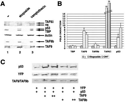
FIG. 6.
TAF9 and TAF9b are differently induced by apoptotic stimuli and involved in p53 stabilization. (A) HeLa cells were treated with etoposide VP16 (Et-VP16; 68 μM) or camptothecin (CMT; 15 μM). Nuclear extracts were prepared after no treatment (−) or the indicated treatment for 6 h and analyzed by SDS-PAGE followed by immunoblotting with the indicated antibodies. The samples were normalized by loading the same amount of actin. ns, nonspecific. (B) Three independent experiments as shown in panel A were densitometrically scanned, and the induction (n-fold) of the expression of different indicated proteins (compared to actin) is represented in the graph. The induction of TAF6δ is indicated on the top of the corresponding bars. Error bars are shown. (C) MCF7 cells were transfected with an expression vector for p53 alone or cotransfected with increasing amounts of TAF9 or TAF9b expression vectors. Transfection efficiency was monitored by cotransfecting pEYFP. Total cell extracts were prepared after 48 h and analyzed by SDS-PAGE followed by immunoblotting for the presence of p53 and YFP. TAF9 and TAF9b were detected with a polyclonal antibody that recognizes both proteins.

