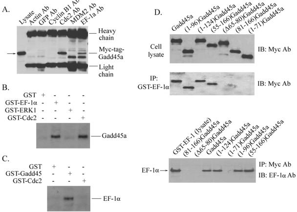
FIG. 6.
Interaction of Gadd45a with EF-1α. (A) Myc-tagged-Gadd45a vector was transiently expressed in HeLa cells via Lipofectamine transfection. At 48 h posttransfection, whole-cell protein extracts were prepared and immunoprecipitated with anti-actin, anti-GFP, anti-Cdc2, anti-MDM2, and anti-EF-1α antibodies. The immunocomplexes were analyzed by sodium dodecyl sulfate-polyacrylamide gel electrophoresis and Western blotting with anti-c-Myc antibody. (B) Whole-cell lysates prepared from HeLa cells were incubated with GST-EF-1α, GST-ERK1, GST-Cdc2, or GST alone (conjugated with Sepharose beads) for 6 h at 4°C. Immunocomplexes were then washed four times with lysis buffer and analyzed with antibody to Gadd45a. (C) GST-Gadd45a, GST-Cdc2, and GST alone were incubated with HeLa cellular protein as described in panel B. Immunoblotting assays were used to examine the presence of EF-1α protein in immunocomplexes by using anti-EF-1α antibody. (D) A series of Myc-tagged-Gadd45a deletion mutants were introduced into HeLa cells via transient transfection. Whole-cell lysates were prepared and incubated with GST-EF-1α (conjugated with Sepharose beads). Immunocomplexes were then washed four times with lysis buffer and analyzed with anti-Myc antibody. In addition, cellular lysates isolated from HeLa cells transfected with a series of Myc-tagged Gadd45a deletion mutants were incubated with GST-EF-1α protein and immunoprecipitated with anti-Myc antibody. Immunoprecipitates were analyzed with anti-EF-1α antibody.

