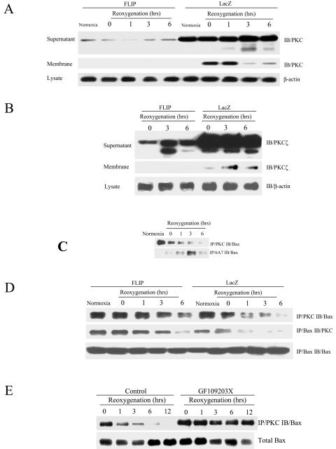
FIG. 4.
PKC is inactivated by FLIP and associates with Bax. MLEC at 30% confluence were cultured in serum-free media in the presence of 106 CPU/ml of adeno-FLIP and adeno-lacZ for 3 h and then restored to normal medium. Two days later, cells were exposed to hypoxia (95% N2, 5% CO2) for 24 h and then restored to normal culture conditions (95% air, 5% CO2) for the indicated times. Cell lysates were separated into membrane and supernatant fractions and subjected to immunoblotting (IB) to detect PKC using total PKC antibody (A3) raised against PKCα (A) or ζ-specific anti-PKC (B). MLEC grown to 90% confluence were exposed to hypoxia (95% N2, 5% CO2) for 24 h and then restored to normal culture conditions (95% air, 5% CO2) for the different periods of time indicated. Cell lysates were subjected to direct immunoprecipitation with PKC (A3) or activated Bax (6A7) antibodies followed by immunoblotting to detect Bax (IB) (C). Total lysates as described for panel A were subjected to direct immunoprecipitation with PKC or Bax followed by immunoblotting to detect Bax or PKC (D). MLEC grown to 90% confluence were exposed to hypoxia (95% N2, 5% CO2) for 24 h in the absence or presence of protein kinase C inhibitor GF109203X (400 ng/ml) and then restored to normal culture conditions (95% air, 5% CO2) for the different periods of time indicated. Cell lysates were subjected to direct immunoprecipitation with PKC antibody (A3) followed by immunoblotting to detect Bax (E).

