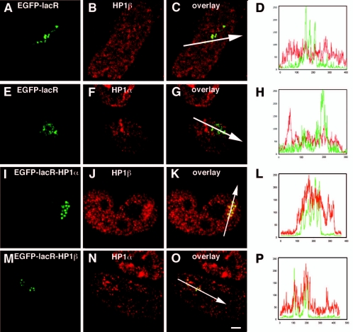
FIG. 7.
HP1 targeting induces the recruitment of endogenous HP1. RRE_B1 cells transfected with EGFP-lacR (A to H), EGFP-lacR-HP1α (I to L), or EGFP-lacR-HP1β (M to P) were, after 48 h, immunofluorescently labeled with an antibody against HP1β (A to D and I to L) or against HP1α (E to H and M to P). 3D images were recorded; the images shown represent individual midnuclear optical sections. The green signal shows the EGFP-lacR-tagged chromosomal array, and the red signal shows the distribution of immunolabeled endogenous HP1. Panels D and H show line scans through the nuclei shown in panels C and G, respectively, and panels L and P show line scans through the nuclei shown in panels K and O, respectively; the position of the line scan is shown by the white arrow. Nuclei in panels A to C, E to G, I to K, and M to O are on the same scale. Bar, 1.4 μm.

