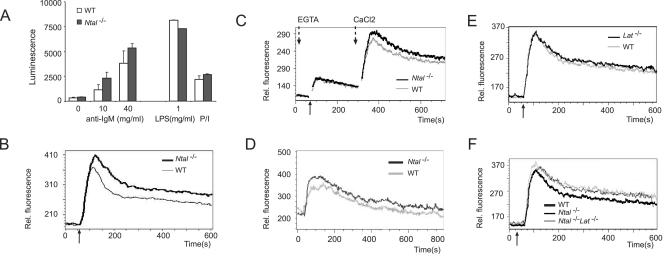
FIG. 5.
Proliferative and Ca2+ responses in splenic B cells from Ntal−/− mice and Lat−/− Ntal−/− double-deficient mice. (A) Spleen B cells purified from wild-type (WT) or Ntal−/− mice were stimulated with increasing concentration of goat F(ab)′2 anti-mouse IgM antibody (0-40 μg/ml), with lipopolysaccharide (1 μg/ml), or with phorbol myristate acetate and ionomycin (P/I). After 40 h of culture, the ATP content of each culture, a value proportional to the extent of cell proliferation, was measured by luminescence. (B) Calcium flux analysis in response to BCR stimulation in wild-type and Ntal−/− mature B cells. The diagram depicts calcium flux (y axis) as a function of time (x axis). Arrow below the x axis indicates the time point of stimulation with F(ab)′2 goat anti-mouse IgM antibody. Data shown are representative of four independent experiments. (C) Intra- and extracellular Ca2+ mobilization was recorded (see Materials and Methods) in mature B cells from wild-type (WT) or Ntal−/− spleens. Same symbols as in panel B. The time point at which the extracellular Ca2+ concentration was restored to 1.3 mM is indicated by an arrow. (D) Calcium flux analysis in response to BCR stimulation in wild-type and Ntal−/− B220+ IgD−, immature B cells. Same symbols as in panel B. (E) Calcium flux analysis in response to BCR stimulation in wild-type, Ntal−/−, and Lat−/− Ntal−/− mature B cells. Same symbols as in panel B. (D) Calcium flux analysis in response to BCR stimulation in wild-type and Lat−/− mature B cells. Same symbols as in panel B.

