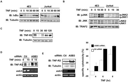
FIG. 9.
ASB3 knockout potentiates TNF-α-induced apoptosis in 4E3 cells. (A) 4E3 and Jurkat cells were treated with TNF-α (10 ng/ml) for the indicated times. Whole-cell extracts were subjected to Western blot analysis with anti-IκBα (top) or anti-β-tubulin (bottom). (B) 4E3 and Jurkat cells were treated as described for panel A. Whole-cell extracts were examined by Western blot analysis with specific antibodies against phospho-JNK (Cell Signaling, Beverly, Mass.), JNK (Pharmingen, San Diego, Calif.), and TRAF2 (Santa Cruz Biotechnology Inc.). (C) Whole-cell extracts were prepared from 4E3 cells following TNF-α treatment for the indicated times. The protein levels of TNF-R2 (top panel) and β-tubulin (bottom panel) were analyzed by Western blot analysis with anti-TNF-R2 and anti-β-tubulin. (D) 4E3 cells, transfected with either control (ctl) siRNA (GFP sequence) or ASB3 siRNA for 48 h, were treated with TNF-α for 15 min. Whole extracts prepared from these cells were subjected to Western blot analysis with anti-TNF-R2. RT-PCRs were performed with the RNAs prepared from these cells to evaluate asb3 expression or β-actin expression. (E) 4E3 cells were transfected with either GFP siRNA (ctl) or ASB3 siRNA for 48 h. Whole-cell extracts prepared from these cells were subjected to Western blot analysis with anti-TNF-R2 and anti-β-tubulin. RT-PCRs were performed with RNAs prepared from these cells to evaluate asb3 expression or β-actin expression. 4E3 cells were transfected with a Renilla luciferase reporter construct (pRLTK; Promega) and either control siRNA or ASB3 siRNA for 48 h. Each group was split in two, with one group maintained in medium and the other treated with TNF-α. At the indicated times, the percentage of specific cytotoxicity was determined by measuring the loss of Renilla luciferase activity. The means and standard deviations of three independent experiments are shown.

