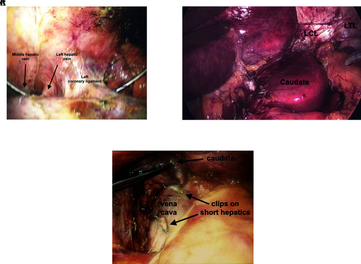
Figure 3.
Division of left coronary ligament. (A) Exposure of left hepatic vein adjacent to left coronary ligament with probe between middle and left hepatic vein. (B) Exposure and division of posterior left triangular (LTL) and coronary ligament (LCL). This allows full visualization of the fissure, as well as can be used to mobilize the and resect the caudate lobe. (C) Caudate lobe can be lifted off vena cava after division of short hepatic veins with laparoscopic regular and micro clips.

