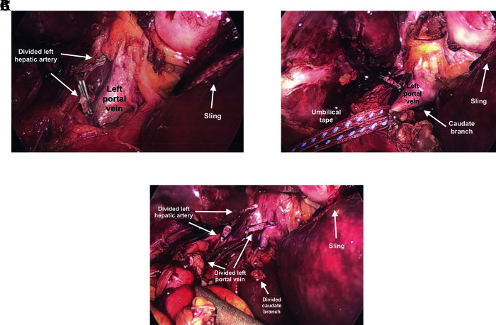
Figure 4.
Laparoscopic hilar dissection for left lobectomy. (A) Divided left hepatic artery and exposure of left portal vein. Sling pulling left lateral segment. Traction on hilar plate allows easier dissection of left portal vein. (B) Umbilical tape applying traction to left portal vein prior to dividing. One needs to be mindful of caudate lobe branch off of left portal vein. (C) Divided left portal vein and caudate lobe branch.

