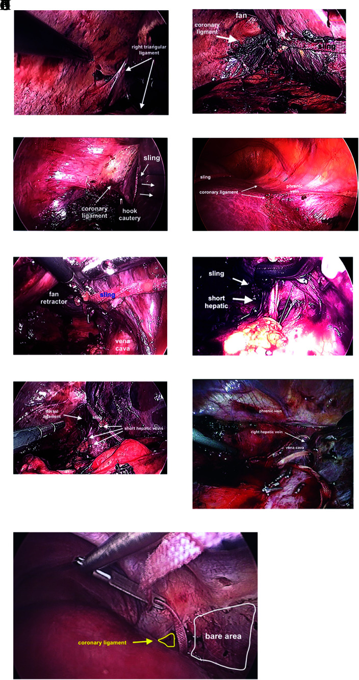
Figure 8.

Mobilization of right lobe. (A) Right triangular ligament prior to dividing with a hook cautery. (B) Liver mobilized to the left facilitated with sling, fan retractor and division of coronary ligament. (C) Dome of liver retracted inferiorly opening coronary ligament aided by sling. (D) Division of anterior coronary ligament aided by retraction of liver to towards left foot aided by sling. (E) The “Batcave,” after coronary ligament divided as much as possible, the right lobe can be lifted off the vena cava aided by a fan retractor and sling allowing visualization the division of short hepatic branches. Care should be taken to prevent the sling from sliding into vena cava and tear a short hepatic vein. (F) Exposure short hepatic vein with right angle clamp. (G) Dorsal hepatic ligament prior to division. (H) Dorsal hepatic ligament after division, allowing full release of right lobe of liver. (I) Golden finger used to feed umbilical tape between right and middle hepatic veins.
