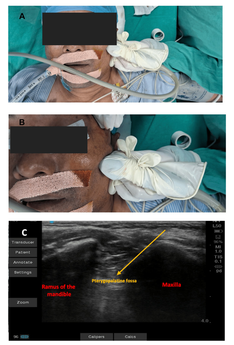
Figure 4. Real-time ultrasound-guided sphenopalatine ganglion blockade.
(A) Placement of linear ultrasound probe inferior and parallel to the zygomatic process. (B) Block needle inserted just behind the posterior orbital rim and above the zygomatic process, directing it 45° caudally. (C) Corresponding ultrasound image to perform the sphenopalatine ganglion block; local anesthetic injected into the pterygopalatine fossa.

