Fig. 6.
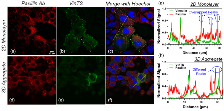
CHO-K1 cells expressing VinTS and immunostained with paxillin primary antibody labeled with Alexa-647-conjugated secondary antibody in a 2D monolayer (top panels) and a 3D multicellular aggregate (bottom panels). (a, d) Alexa-647 fluorescence (red pseudocolor), (b, e) VinTS fluorescence (green pseudocolor). (c, f) Merged images including Hoechst staining of the nuclei (blue pseudocolor). (g) and (h) Signal cross sections along the lines in the merged images (c and f) show overlapping peaks in the Alexa-647 and VinTS channels at sites of VinTS expression in the 2D monoloayer but separate peaks in the 3D multicellular aggregate.

