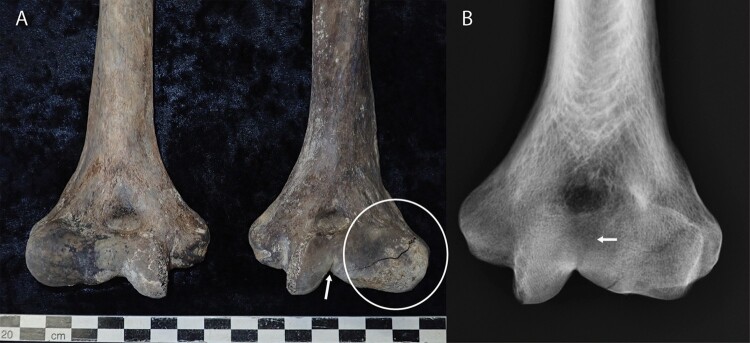
Figure 7.
Distal humerii of the Burial 11 (A). A depressed linear defect is observable on in apex of the trochlear space (White arrow) and extends to the posterior trochlear surface. The capitulum of the left humerus (white circle) is distorted laterally and inferiorly. The capitululum is considerably enlarged compared the right hand side (pictured left) which is the normal rounded shape for articulation with the radial head. The distortion of the capitulum is clinically described as a ‘fishtail deformity’. B, Radiograph of left distal humerus. The white arrow shows the remains of the fracture line extending from the linear defect in the trochlear apex. This injury likely occurred during childhood (<14–19 years).

