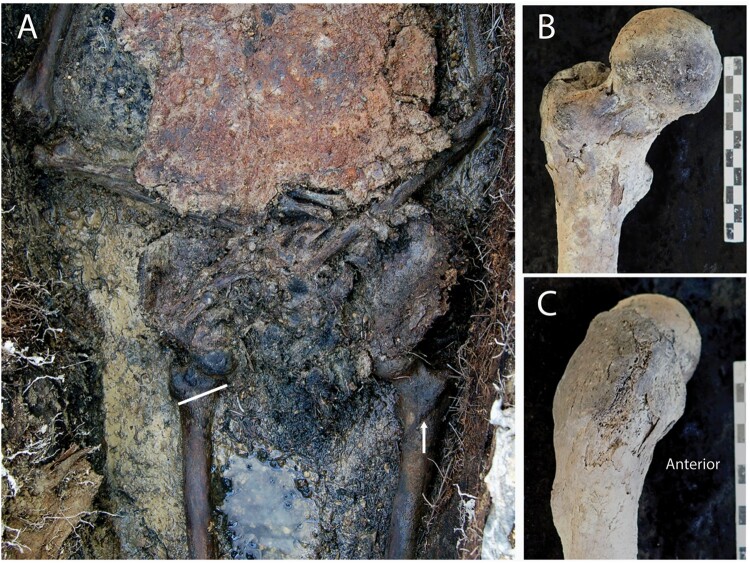
Figure 10.
The right femur of Burial 13. A, In situ image of the pelvis and hip area showing the transverse (abnormal) orientation of the right intertrochanteric line (attachment for the iliofemoral ligament of the hip joint) compared with the oblique (normal) orientation on the left side. B and C, The proximal third of the right femur is significantly bowed anteriorly. No fracture callus was appreciated in radiography of the bone.

