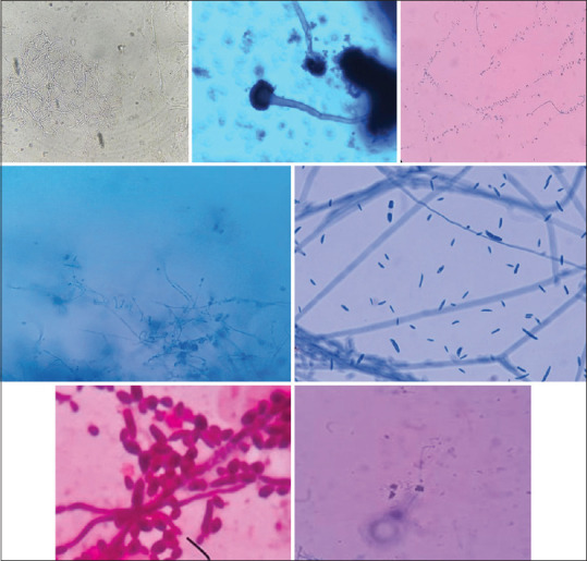
Figure 1.

Microscopic images of: (a) KOH wet mount showing thin,long fungal hyphae (b) Aspergillus fumigatus species, LPCB (40×) (c) Tear drop shaped microconidia of T.rubrum, LBCB mount (d) Culture of microconidia and spiral hyphae of T.mentagrophytes, LPCB (40×) (e) Sickle shaped macroconidia suggestive of Fusarium species, LPCB mount (40×), (f) Round to oval budding yeast cells with pseudohyphae suggestive of C.albicans (100×) (g) Clusters of microconidia and spiral hyphae of T.mentagrophytes, LBCB mount (40×)
