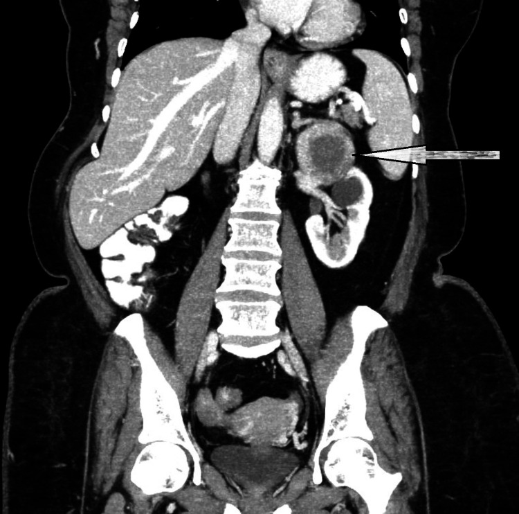
Figure 3. Coronal CT of abdomen and pelvis.
A large well-defined hypoattenuating, non-enhancing cystic lesion of size 71 x 61 x 56 mm, surrounded by enhancing adrenal tissue was noted arising from the inferolateral aspect of the left adrenal gland, seen abutting the tail of the pancreas and the small bowel loop (arrow). It showed no solid components/septations/calcifications.

