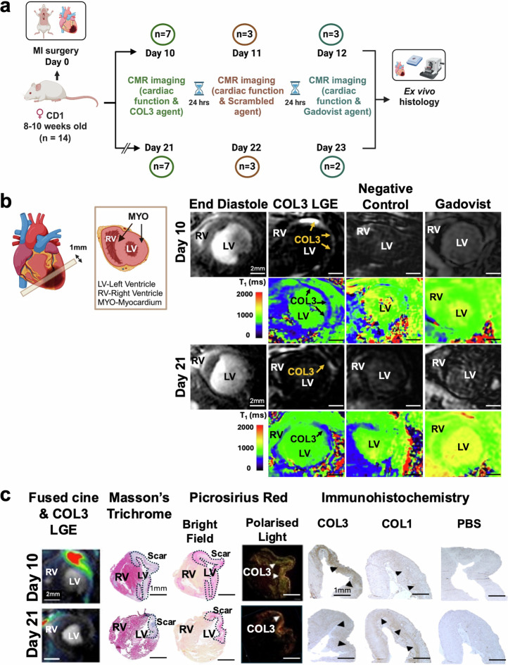
Fig. 5. First in vivo molecular imaging of COL3 after myocardial infarction using the tetrameric [Gd-DOTAMA]4-CBP1 probe.
a Experimental design. b Molecular CMR (at 60 min post-injection of the COL3 probe) shows strong signal enhancement and lower T1 values in the infarcted myocardium at day 10. Conversely, low signal enhancement and higher T1 values are observed in the infarct at day 21. No enhancement is observed with both the negative control probe and the non-targeted clinical probe (Gadovist) (at 30 minutes post-injection). c MRI signal enhancement co-localised with collagen seen on Masson’s trichrome and PSR staining in BF and with COL3 fibres (green fibres) seen with PLM. Immunohistochemistry validates co-localisation of the MRI signal with COL3 at day 10 and the drop in MRI signal at day 21 when COL3 is replaced by COL1. RV right ventricle, LV left ventricle, PSR Picrosirius Red, BF bright field, PLM polarised light microscopy. Figure 5a and schematic in 5b were created with BioRender.com.

