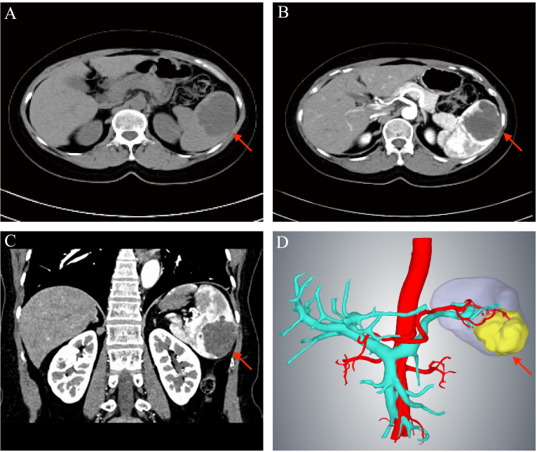
Figure 1.
Preoperative examination of the patient. (A) Computed tomography scan showing a mass of about 61×15 mm in the lower pole of the spleen (red arrow). (B) Enhancement of the wall of the capsule with no obvious enhancement within the capsule is observed (red arrow). (C) The red arrow indicated the tumor. (D) Three-dimensional imaging showing the tumor confined to the lower pole of the spleen (red arrow) and supplied by the lower pole splenic artery.

