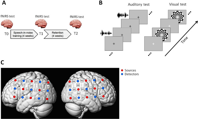
Fig. 2.
Experiment design. (A) Participants completed a 4-week home-based speech-in-noise training and their brain functions were measured by fNIRS before (T0) and after the training (T1 and T2). (B) The fNIRS experiment included an auditory test where participants listened to auditory sentences (speech and spectrally rotated speech) and a visual test where participants watched a flashing chequerboard. A block design was adopted with resting blocks interleaved between the auditory/visual stimuli. (C) Optode configuration of the fNIRS experiment was two 5-by-3 probe sets that formed 44 channels (22 channels in each hemisphere) over speech- and language-related temporal, parietal and frontal cortical regions (left: left hemisphere; right: right hemisphere). Red and blue circles denote the sources and detectors, respectively. The channel indices are indicated in the white squares between the sources and detectors

