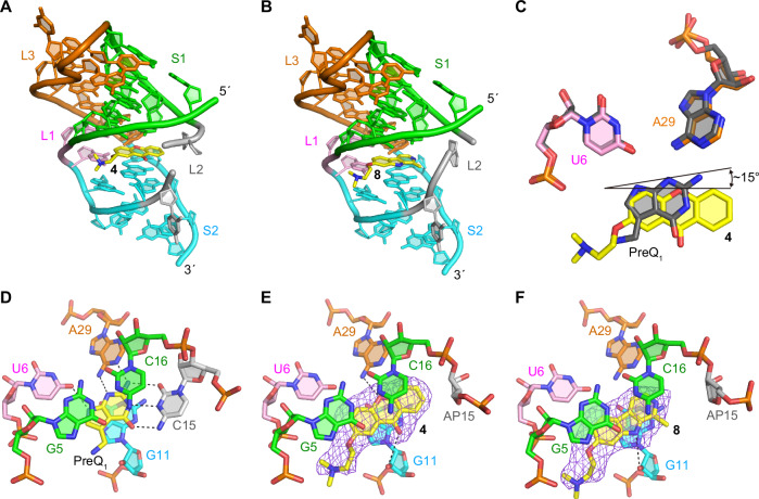
Fig. 2. X-ray crystal structures of ab13_14_15 in complex with synthetic ligands.
A, B Overall structure of the complexes with ligands 4 (A) and 8 (middle). C Comparison of binding poses between 4 and PreQ1. D Structural comparison between the wild-type Tte-PreQ1 riboswitch aptamer complexed with PreQ1 (PDB ID: 3Q50)70, (E) ab13_14_15 complexed with 4 and (F) ab13_14_15 in complex with 8. Hydrogen bonds are shown in dashed lines. Purple mesh represents the mFo-DFc electron density maps observed for each ligand, which are contoured at 2.5 σ. The compounds were omitted from the phase calculation. The distances between the oxygen atom of the central ring of 4 and the N6 atom of A29, the carbonyl group of 4 and 2’-OH group of G11, and the amine of the pyrrole ring of 8 and 2’-hydroxyl group of G11 are 3.63, 3.75, and 3.66 Å, respectively.

