. 2024 Sep 2;24(20):4816–4826. doi: 10.1039/d4lc00547c

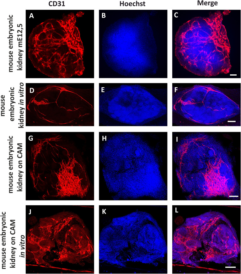
Fig. 7. Detection of the blood vessels in mouse embryonic kidneys in different variants of the experiment. Positive control: intact mouse embryonic kidney dissected from E12,5 embryo (A–C). Negative control: mouse embryonic kidney dissected from E12,5 embryo, cultivated 5 days in vitro (D–F). In vivo experiment. Mouse embryonic kidney E12,5 was vascularized on a chicken embryo CAM for 4–5 days (G–I). In vivo experiment after continuation in vitro. Mouse embryonic kidney E12,5 was vascularized on the chicken CAM for 4–5 days, and then cultivated 5 days under in vitro conditions (J–L). CD31 was used as an endothelial marker (red) and Hoechst as a nuclear marker (blue). Scale bars = 50 μm.

Fig. 7