Figure 1.
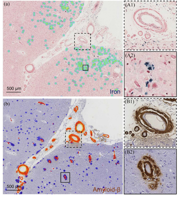
Examples of iron deposits in the cortex, cortical and leptomeningeal CAA and leptomeningeal grade III vessel remodeling. Examples of the application of a deep learning model (Aiforia®) for iron (a) and amyloid (b) in two adjacent brain sections of a participant with CAA. (a) Cortical iron deposits recognized by the model are overlaid in green (stained blue, see corresponding inset A2). (b) Parenchymal amyloid-β recognized by the model is overlaid in blue, CAA (cortical and leptomeningeal) recognized by the model in red (see corresponding inset B1 and B2, amyloid-β stained brown). Inset A1 and B1 shows an example of a single leptomeningeal vessel with Vonsattel grade III remodeling (vessel-in-vessel pathology). The inset B2 shows an example of a cortical vessel with dyshoric CAA.

