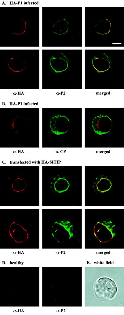
FIG. 6.
Analysis of the colocalization of P1, P2, and CP in AMV-infected cowpea protoplasts by immunofluorescence microscopy. Each column of images shows the analysis of a single protoplast: left and middle, protoplasts analyzed with specific antisera; right, the left and middle images merged. (A) Two protoplasts infected with AMV that was engineered to express HA-tagged P1. The protoplasts were analyzed with antisera against the HA epitope (left) and P2 (middle). (B) Protoplast infected with AMV that was engineered to express HA-tagged P1. The protoplast was analyzed with antisera against the HA epitope (left) and CP (middle). (C) Two protoplasts transfected with a plasmid expressing γ-TIP fused to an HA epitope. The protoplasts were analyzed with antisera against HA (left) and P2 (middle). (D) Healthy protoplasts analyzed with antisera against the HA epitope (left) and P2 (middle). (E) White-field picture of the lower protoplast shown in panel C. Bar = 10 μm.

