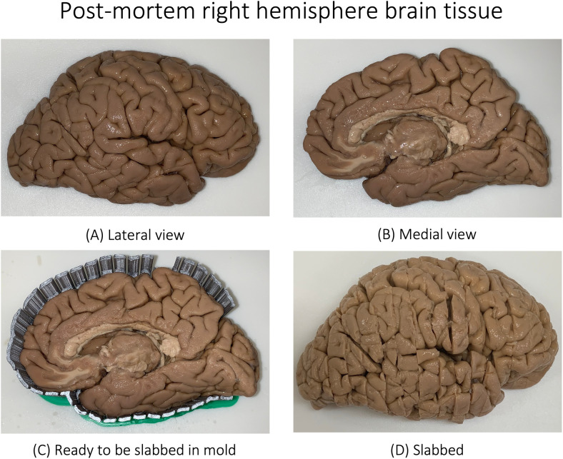
Fig. 1.
Postmortem tissue blockface photograph of a donor with diagnosis of Parkinson’s disease (not demented) and Lewy body disease (deceased at the age of 79). Shown are the lateral (A) and medial (B) views of the right hemisphere. The tissue is then placed in a mold (C) and is subsequently slabbed (cut into different slices) as shown (D). See Lasserve et al., 2020 for more details. See Supplementary Figure 1 for the slices of the given brain tissue.

