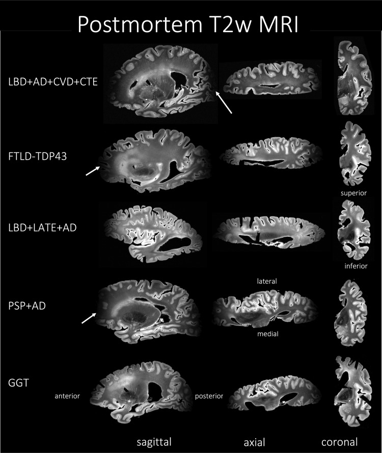
Fig. 2.
MRI of the T2w sequence representative of Alzheimer’s disease and related dementias (ADRD) spectrum with mixed pathology and diagnoses of five subjects. The heterogeneity among the subjects can be appreciated through the three different viewing planes. Notice that the MRI signal drops off at the anterior and posterior ends of the hemisphere, a drawback of the current acquisition protocol. AD, Alzheimer’s disease; CVD, cerebrovascular disease; LATE, limbic-predominant age-related TDP-43 encephalopathy; LBD, Lewy body disease; CTE, chronic traumatic encephalopathy; FTLD-TDP, frontotemporal lobar degeneration with TDP inclusions; GGT, globular glial tauopathy; PART, primary age-related tauopathy; PSP, progressive supranuclear palsy.

