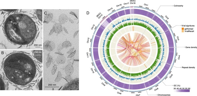
Figure 1.
Morphologic and genomic characteristics of the Bathycoccus sp. UST710. (A, B) Transmission electron microscopy (TEM) images of Bathycoccus sp. UST710 cells revealing the nucleus (N), single chloroplast (C), mitochondrion (M), vesicles (V), starch grain (SG), plastoglobuli (PG), and scales (S) covering the cell surface. Scale bars: 200 nm. (C) TEM image displaying a detailed view of the scales. Scale bars: 200 nm. (D) Physical map of the genome highlighting the key features of this isolate. The outermost track illustrates the size of 18 chromosomes, labeled Chr1-18 in descending order of size, with two outlier chromosomes—the BOC and the SOC—labeled and highlighted. Proceeding inward, four tracks represent the distribution of GC content (5-kb sliding windows), repeat element density (10-kb sliding windows), gene density (10-kb sliding windows), and predicted viral regions identified by geNomad and ViralRecall. Syntenic gene blocks, identified by MCScanX, are connected by links at the center.

