FIG. 5.
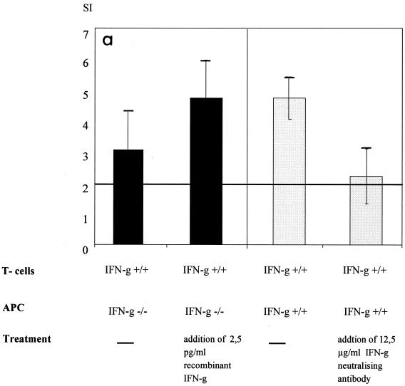
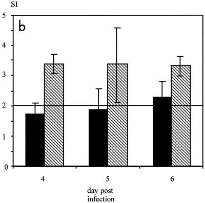
Neutralization of IFN-γ inhibits antigen processing and T-cell proliferation in vitro and in vivo. (a) MV-specific CD4 T cells were stimulated with UV-inactivated MV and IFN-γ+/+ or IFN-γ−/− APC. In addition, either recombinant IFN-γ (2.5 pg/ml) or IFN-γ-neutralizing antibodies (12.5 μg/ml) were added. The differences in the SI with and without the addition of recombinant IFN-γ were significant (P < 0.05, Kruskal-Wallis test), as were the differences with and without neutralizing antibody (P < 0.05, t test) (error bars indicate standard deviations). The lack of IFN-γ did not reduce constitutive MHC-II expression on APC, whereas the addition of IFN-γ increased it (data not shown). Concanavalin A-dependent proliferation was not influenced by the lack or addition of IFN-γ (data not shown). In addition, concanavalin A-dependent proliferation of spleen cells from naive animals was not influenced by IFN-γ concentrations ranging from 40 ng to 0.04 pg/ml (data not shown). (b) BALB/c mice were infected i.c. with 5 × 10⁴ TCID50 of MV strain CAM/RBH and injected daily with IFN-γ-neutralizing antibodies (black bars) or left untreated (striped bars). On days 4, 5, and 6, MV-specific T-cell proliferation was measured from spleen cells (error bars indicate standard deviations). The levels of proliferation of spleen cells from these animals after concanavalin A stimulation were comparable (data not shown).


