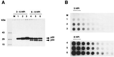
FIG. 2.
(A) Expression of early (p30) and late (p54) ASFV proteins in infected macrophage cell cultures. Immunoprecipitation of cell extracts from mock-infected macrophages (lane M) and macrophages infected with E70 (lanes 1 and 4), MS16 (lanes 2 and 5), or BA71V (lanes 3 and 6), radiolabeled from 3 to 6 or 6 to 9 hpi, was performed using a mixture of anti-p30 and anti-p54 monospecific rabbit antiserum as previously described (1). Sizes were estimated using Rainbow 14C-methylated protein molecular size markers (Amersham Life Science). (B) Viral DNA replication in E70 (lanes 1 and 4)-, MS16 (lanes 2 and 5)-, or BA71V (lanes 3 and 6)-infected or mock-infected (lane M) macrophage cell cultures. Cells were infected (MOI = 5), total cellular low-molecular-weight DNA was isolated at the indicated times postinfection, and twofold dilution sets of 10 μg of total DNA were blotted onto Zeta Probe membranes (Bio-Rad) and probed with a 32P-labeled E70 genomic DNA probe.

