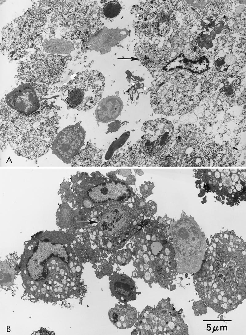
FIG. 3.
Electron micrographs of ASFV-infected swine macrophages. Cell cultures were infected (MOI = 5) with MS16 (A) or E70 (B) virus and examined at 16 hpi. Note the extensive cytopathology in MS16-infected macrophages compared to those infected with E70. Virus factories (arrows) are present in the cell cytoplasm.

