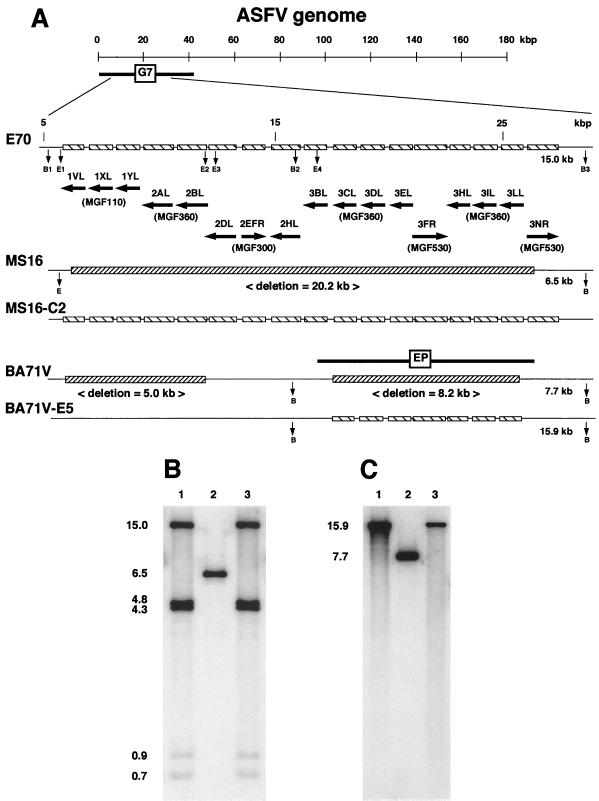
FIG. 6.
Characterization of marker-rescued MS16-C2 and BA71V-E5 viruses. (A) Diagram of the left variable region in ASFV pathogenic isolate E70, cell culture-adapted MS16 and BA71V viruses, and rescued recombinant viruses MS16-C2 and BA71V-E5. (B and C) Southern blot analysis of E70 (lanes B1 and C1), MS16 (B, lane 2), MS16-C2 (B, lane 3), BA71V (C, lane 2), and BA71V-E5 (C, lane 3). Purified viral DNAs were digested with EcoRI/BamHI (B) or BamHI (C), electrophoresed, blotted, and hybridized with DNA probes including the deleted regions of MS16 and BA71V. Positions of molecular size markers are shown in kilobase pairs at the left.

