



Abstract
Regeneration is a heroic biological process that restores tissue architecture and function in the face of day-to-day cell loss or the aftershock of massive injury. Capacities and mechanisms for regeneration can vary widely among species, organs, and injury contexts. Here, we describe hallmarks of regeneration found in diverse settings of the animal kingdom, including activation of a cell source, initiation of regenerative programs in the source, interplay with supporting cell types, and control of tissue size and function. We discuss these hallmarks with an eye toward major challenges and applications of regenerative biology.
Short summary:
Poss and Tanaka provide a review of concepts and mechanisms in regenerative biology. They describe “hallmarks” that are broadly represented across species, tissues, and injury contexts, as a means to articulate structure for addressing knowledge gaps and potential applications.
Introduction
Regeneration, the restoration of lost cells, tissues, body parts, or whole animals, is a central feature of multicellular life. Development from embryonic to adult stages incurs cell death, stresses, and strains that provoke and guide morphogenesis, events akin to regeneration. Final form is not truly final -- cycles of renewal maintain critical tissues, such as blood and epithelial linings.
Mammals are vulnerable to heart attacks, spinal cord injury, limb amputation, and stroke, for which regenerative responses are inadequate. Molecular, cellular, and tissue damages accumulate as animals age, and the ability to resolve these insults is tempered. Regeneration can counter these damages and bring near-immortality to some species, such as flatworms and polyps. Other vertebrate species, among them salamanders and teleosts, show extraordinary capabilities to regenerate limbs, hearts, spinal cord, and more. Yet, the defense mechanism of regeneration inevitably falls short in vertebrates. Aging conquers regeneration in all scenarios.
Regeneration is in some ways similar to cancer, and attempts at comparisons have been frequent over the past decades. Most notably, each process commandeers the machinery of cell growth and patterning for its purposes. However, some may consider these two players to be diametrically opposed. DNA-based changes underlie cancer, and cancer cells eventually identify cheat codes to escape checks and balances, with insidious outcomes. In contrast, regeneration is initiated by an injury event, with an ostensible goal to achieve perfect form and/or function. Therapies to improve human regeneration are an inspirational objective of the 21st century. Yet, both a hammer and scalpel are needed to launch morphogenesis in a controlled manner from a disabled organ or tissue stump, avoiding tumorigenesis and metastasis.
Regeneration is an historic and decorated field of biology, and many laboratory model systems are employed with their own spectra of regenerative capabilities. Discoveries in this field can inspire regenerative medicine. Here, we attempt to articulate a simple framework for understanding regeneration. Our aim is broad, and we skew our examples toward non-mammalian species, such as planarians, salamanders, fish, and the like – laboratory model systems with which many readers might wish to gain more familiarity. Principles of tissue organization, molecular pathways, and injury responses are conserved across phylogeny, and species with outsized regenerative abilities can serve as role models for mammals toward a potential explosion of the field if methodologies allow. We describe four “hallmarks” of regeneration that typically occur in response to acute trauma, covering what we have learned and must strive to learn about their defining features (Fig 1). Upon damage and initial healing, a competent cellular source(s) of lost tissues must be activated, programs for morphogenesis are initiated within and upon this source, productive cell interactions occur, and new and old cells functionally integrate and restore size and shape.
Fig 1.
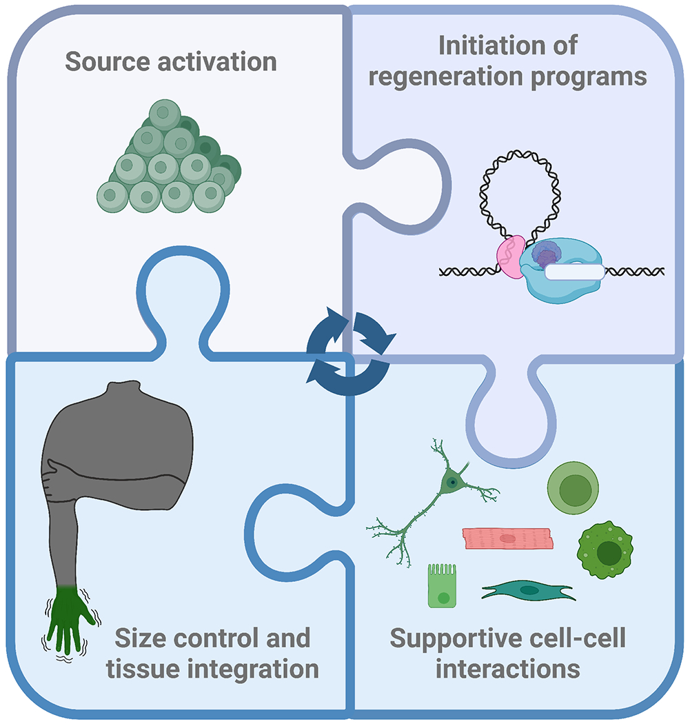
Hallmarks of regeneration.
1. Activation of cell sources
Trauma leads to an initial healing response of some form in all tissues, which can involve events such as clotting and re-epithelialization. When healing is complete in a regeneration-competent tissue, renewal of structure and function begins. A complex tissue has many cell types, and each may have a different origin and/or mechanism of origin. A reserve of stem cells may be involved, or differentiated cells might be tweaked in a manner that relieves them of function and enables division. The lesson learned from experiments across many body parts, injury types, and animal species is that every possible way of recruiting source cells for regeneration – stem cell activation, dedifferentiation, transdifferentiation, hypertrophy and endoreplication – has been observed.
Stem cells.
Stem cells can self-renew and give rise to other cell types. Arguably the most familiar and elegant starter material for regeneration is the stem cell and its requisite niche (Fig 2A). Cell loss or tissue damage signals disrupt the serenity of the niche, and stem cells awaken tasked to restore function. Many classic and exceptionally well-studied examples of stem cell-based regeneration have been best studied in mammalian models, such as mice. These include blood, derived from hematopoietic stem cells in bone marrow niches, and skeletal muscle, derived from satellite cells located beneath the basal lamina of multinucleated myofibers. Skin and its appendages contain dynamic stem cell populations that regenerate the epithelial barrier (Fig 2B), as well as hair growth and color1–3.
Fig 2. Activation of cell sources.

A. Stem cells reside in a quiescence-maintaining niche before they wake to renew and differentiate upon cell loss or damage.
B. Dynamic stem cell populations in the skin regenerate the epithelial barrier.
C. Pluripotent stem cells populate interorgan spaces in planarian flatworms and replace differentiated cells in the process of turnover.
D. Injury-induced partial dedifferentiation of cardiomyocytes leads to acquisition of embryonic features and division during zebrafish heart regeneration.
E. Iris pigmented epithelial cells transdifferentiate to lens cells in amphibians, by depigmenting, forming a vesicle, and initiating production of lens crystallin proteins.
F. Spared Drosophila pyloric cells endocycle in response to genetic ablation, forming a structure composed of a reduced number of large, polyploid cells with effective barrier function.
Other spectacular examples of regeneration involve pluripotent stem cell populations, for example in planarian flatworms (Fig 2C). Stem cells in the form of neoblasts populate interorgan spaces of planarians and mediate turnover. Some of these neoblasts are pluripotent, demonstrated by the ability of single transplanted cells to repopulate multiple tissue types and even save an animal from lethal irradiation4. Neoblasts have been classified by morphology and transcriptomes, and the surface marker TSPAN-1 emerged as an indicator of a pluripotent class5, with other pluripotent classes likely present6. Certain other neoblast classes are more committed to fates of the various tissues, for instance intestine, muscle, nervous system, or pharynx7. Key research goals for this system, and for virtually all stem cell-based systems, include further detailing the stem cell hierarchy and the signals and steps toward fate commitment, and determining how neoblasts sense the extent of injuries and adjudicate their response. Contemporary studies of stem cell biology have used multiplexed clonal analysis and barcoding approaches to tag related cells and track them animal-wide. Methods for transgenesis that enable direct, live visualization of neoblasts and their progeny in species such as acoel worms is an important future direction to complement inferential methods8.
Direct duplication and dedifferentiation.
Many tissues regenerate without using stem cells by duplicating parenchymal cells, and dedifferentiation is a process that can assist in this. In innate regenerative biology, dedifferentiation typically refers to the tempering of functional properties of a specialized adult cell. This process can involve the reduced expression of signature “differentiation” genes of that cell and/or increased presence of a more “embryonic” set of genes that is associated with the growth and function of the organ’s initial form. Many examples of dedifferentiation are evident in innate regeneration. For instance, zebrafish and other non-mammalian vertebrates can regenerate heart muscle after extreme loss through injury-activated division of cardiomyocytes9 (Fig. 1D). During zebrafish heart regeneration, transcriptomes of regenerating cardiomyocytes acquire similarities with embryonic cardiomyocytes, including expression of transcription factors and myosin forms associated with heart development, while also dissembling sarcomeres and acquiring a more glycolytic (versus oxidative) state10,11. Importantly, hearts of neonatal mice and other mammalian neonates also display a regenerative response to injury, with limited scarring as an adult mammal would display12–14. Neonatal heart regeneration also features cardiomyocyte division, dedifferentiation, and acquisition of glycolytic features14–16.
Regeneration of an amputated salamander limb involves blood loss, major skin damage, and depletion of large regions of muscle and skeletal bone or cartilage. Thus, to achieve regeneration, the animal effectively requires everything, everywhere, all at once. Although each of these tissues has its own progenitor mechanism, cell source mechanisms for a specific tissue can vary among species, even those in the same vertebrate class or order, and depend on the developmental stage of the animal. Newts and axolotl limb regeneration each replenish limb skeletal muscle after amputation injury. Larval axolotls and newts rely on satellite cells for skeletal muscle regeneration, as do postmetamorphic axolotls, based on cell transplantation and genetic fate-mapping assays17,18. However, adult newts do not rely on satellite cells during limb regeneration, but instead on a process of dedifferentiation18. Differentiated myofibers undergo fragmentation into a mononuclear cell population that regenerates lost skeletal muscle.
In mammalian lungs, airways and alveoli contain stem cells that promote vigorous renewal of compartment-specific epithelial cell types in response to inhaled insults19. Many genetic tools now exist to ablate specific cell populations in these structures, including the major stem cell and differentiated cell types, and studies employing these have revealed notable plasticity among lineages. For example, induced, extreme genetic ablation of large airway basal cells, by targeted diphtheria toxin A expression via a tamoxifen-regulated Cre transgene, enables dedifferentiation of detoxifying club cells to form multipotent basal cells that respond to additional injury20.
Dedifferentiation has been an attractive target for changing regenerative capacity. Forced expression of classic reprogramming factors can affect the chromatin status and transcriptional readouts of differentiated cells, reverting cells at low frequency to a pluripotent state. Multiple recent studies have reported that transient expression of reprogramming factors (Oct4, Sox2, Klf4 and c-Myc; OSKM) alter the regenerative responses of mammalian tissues that normally display poor regenerative capacity21–24. The effects of this type of partial reprogramming have been examined in many tissues, and have been reported, for instance, to improve the ability of mouse cardiomyocytes to divide and respond to injury stimuli21–24. The concept of partial reprogramming brings interesting ideas to regenerative biology, although the tumorigenic effects of OSKM factors make it difficult to imagine therapeutic use without exceedingly sophisticated safety measures. Other manipulations have reported pro-regenerative effects of transcription factor overexpression on the ability of tissues, such as skeletal or heart muscle, to dedifferentiate25,26. It is important to catalog manipulations that toggle differentiation in productive ways.
Transdifferentiation and ancillary sources.
Transdifferentiation is the transition from one differentiated cell type to another, and for the purpose of this review we can distinguish transdifferentiation from reprogramming as an event not involving experimentally forced expression of exogenous genes. Transdifferentiation is related to dedifferentiation in that it can involve (but does not require) progression of a cell through an undifferentiated state such as a stem cell. While a dedifferentiated cell remains undifferentiated or redifferentiates to its previous cell type, transdifferentiation takes the trajectory on a new path to a different cell type.
Prominent examples of innate transdifferentiation have been discovered in studies of regeneration of eye parts, which occurs effectively in nonmammalian models. For many years the archetype of transdifferentiation has been the lens of the eye in amphibians, such as species of newts. This tissue faithfully regenerates after complete extirpation in adult newts, involving a process of budding from the pigmented epithelial cells of the ventral portion of the iris27. These cells depigment, form a vesicle, and begin to produce lens crystallin proteins to finalize the structure (Fig 2E). This phenomenon can be modeled ex vivo and also occurs in larval axolotls and tadpoles, although in these cases with origins of ventral or dorsal iris, and cornea, respectively28–30. The eye retinae also regenerate to great extents in fish and amphibians after injuries, such as removal, photoablation or neurotoxic damage, occurring also by transdifferentiation 31. In salamanders and tadpoles, the retinal pigmented epithelium transdifferentiates to generate neurogenic precursors for all key cell types 31,32, whereas structural Müller glial cells distributed throughout the retina repopulate neuron classes in teleosts, such as zebrafish33.
Mammalian pancreas, stomach, and liver are capable of high degrees of transdifferentiation, but only in response to extreme cell disabling or depletion34–36. Pancreatic islet beta cells have a limited capacity to divide in adult mammals and a poor proliferative response to injuries, such as streptozotocin-induced depletion. Yet, at a low rate and following near total genetic ablation of beta cells in mice, alpha or gamma cells transdifferentiate to acquire beta-cell features such as insulin production, in part through the release of inhibitory signals within islets34,37,38. The digestive chief cells of the stomach undergo metaplasia to gland-like cells upon extreme injury35. Liver regeneration invokes a classic model of direct duplication, wherein hepatocytes lost from a major resection injury will be replaced by division and/or hypertrophy of spared hepatocytes to replace lost tissue mass36,39. However, liver cells show plasticity during regeneration when key cell types are genetically disabled. Hepatocytes can transdifferentiate to bile duct structures after genetic disruptions in Notch signaling perturb biliary development, events potentiated by Tgf signaling40. Conversely, biliary epithelial cells initiate hepatocyte production under various conditions in which hepatocytes are burdened by genetic or toxic insults40–43. Examples of mammalian transdifferentiation may be rare, but they afford inspirational glimpses of the potential of differentiated cells to repurpose themselves in vivo under selective pressure.
Endoreplication and hypertrophy.
Whereas most definitions of regeneration invoke the recovery of cell number as a means for restoring tissue mass, prominent examples of regeneration occur without hyperplasia playing a major role. Mammalian skeletal muscle regeneration, for instance, recovers muscle mass after a lesion through fusion of myoblasts generated from satellite cells, building new multinucleated myofibers and contributing new myonuclei to existing fibers. Dedicated fusogens expressed on each participating cell are responsible for the specificity of these fusion activities, eliminating the possibility of fusion with non-muscle tissue44,45. Liver regeneration, which occurs through different source mechanisms, will in the least severe instances of tissue loss involve endoreplication to duplicate the genome and cause growth of hepatocytes, with classic cell division events being less frequent36,39. This process may entail the formation of mononucleated or multinucleated polyploid hepatocytes, typically with larger cell volumes, derived either from diploid or polyploid hepatocytes. Functions of polyploid hepatocytes are measurably similar to diploid counterparts46; thus, liver regeneration achieves functional goals although tissue architecture and cellularity may differ from the original.
Many mammalian cell types exist with elaborate structural and functional requirements, or restricted capacity for cytokinesis, such as cardiomyocytes, neurons, and chondrocytes. Cell cycle blocks limit the ability of cardiomyocytes to divide in postnatal mammals47–49. Thus, mammalian cardiomyocytes often endoreplicate to increase cell and organ mass during animal growth. Polyploidy further limits the ability of cardiomyocytes to divide after an injury such as an ischemic infarct50,51. This result is also substantiated by findings in zebrafish, in which experimental polyploidization of cardiomyocytes could inhibit injury-induced heart regeneration52.
Additional examples of injury-induced polyploidy have emerged in recent years that highlight benefits of a rapid mechanism to increase cell surface area. In Drosophila, a puncture wound to the abdominal epithelium leads to a combined response of endoreplication and fusion by epithelial cells surrounding the wound53. These enlarged, polyploid epithelial cells efficiently heal large injuries. Flies display an analogous endocycling response when most pyloric cells of the hindgut are genetically destroyed, leading to formation of a pylorus with a reduced number of large, polyploid cells with effective barrier function54,55 (Fig 2F). The recognition of polyploidy as a strategy for tissue regeneration is growing, and genetic and/or in vivo imaging tools such as those readily available in Drosophila should be informative for understanding how and why polyploidy occurs.
2. Initiation of regeneration programs
A simple equation for regeneration requires a competent cell source and an environment with signals to control its morphogenesis. Achieving this competency and environment seems far from simple, however. Thousands of genes sharply alter expression levels during regeneration, either elevating or declining, to effect change in participating cell types. Diverse signaling centers form from spared tissue and dying cells release influential factors. Gene expression levels change in impactful ways away from the injured tissues as well, and previous injuries can prime tissue for regeneration, invoking alerted stages and memory-based responses. It is a great challenge to interpret all inputs and variables.
Regeneration regulatory elements.
Many studies have now profiled chromatin-based indicators of active gene regulation, such as reduced nucleosome occupancy or increased presence of histone modifications, in a variety of species, cell types, and injury contexts56–64. These studies indicate widespread changes in chromatin structure affiliated with regeneration, findings that are complemented by reports indicating roles for chromatin regulatory factors during regeneration65,66. Thus, injury prepares the genome to institute changes in gene expression that enable the release of pro-regenerative stimuli, and the competency of multifarious cells to respond to these stimuli.
Transcriptional responses are afforded specificity by enhancer regulatory elements, during all life stages as well as disease. Regeneration programs are similarly controlled by contextual enhancers, whose sequences likely represent many of the genomic locations of the afore-mentioned changes in chromatin structure. These sequences, called tissue regeneration enhancer elements (TREEs) or similar, can in combination with a permissive promoter direct transient expression preferentially or specifically to regenerating tissue67,68. Candidate TREEs from genomic profiling have been experimentally validated in many regeneration contexts. Whereas mechanosensation of injury might be all or part of the stimuli for transcriptional activation of some TREEs, evidence indicates that other TREEs receive control well after initial injury stress by additional or alternative aspects of regeneration such as inflammation, mitogens, hormones, and patterning factors69, 57. Finding rules for TREEs and what binds them is essential and challenging given the complex in vivo context, and much remains to be discovered. Mutagenesis of enhancer transgenes has revealed regulation in part by common transcription factors, such as AP-1 and TEAD, in zebrafish fin and heart regeneration59,67,70 (Fig 3A). Egr-mediated activation of regeneration-associated genes was demonstrated by bioinformatical predictions and gene knockdown in acoel worms62, among other examples. Silencers that reduce gene expression from homeostatic levels during regeneration can be more challenging to find and verify. Yet, a small number of studies indicate the presence of repressive modules within regulatory elements71,72, or of bona fide silencers that form context-specific interactions with nearby genes and reduce their expression73.
Fig 3. Initiation of regeneration programs.
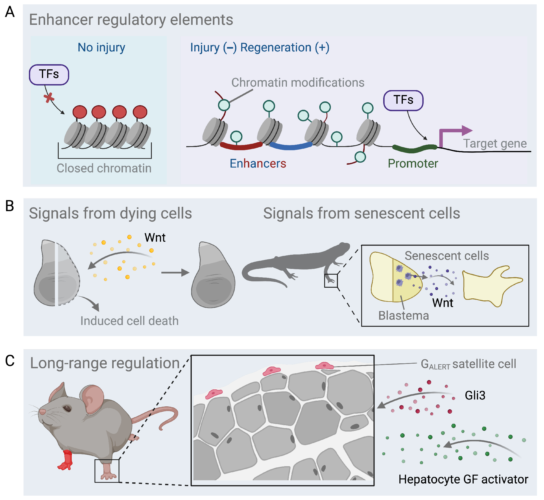
A. Tissue regeneration enhancer elements (TREEs) control the expression of genes induced after injury and during regeneration.
B. Dying Drosophila wing disc cells secrete Wingless (Wnt) to promote compensatory growth of the surviving tissue. Senescent cells transiently appear in the salamander limb blastema and produce mitogenic Wnt ligand.
C. Skeletal muscle satellite cells in one limb respond to a contralateral limb injury by entering a Galert stage, reported to be controlled by circulating Hepatocyte growth factor activator and requiring signaling through the Hedgehog effector Gli3.
As enhancers change rapidly during evolution, they may be targets for evolutionary changes in regenerative capacity59,74. This is an interesting concept that requires further exploration of interspecies differences, but many examples exist in which a limited number of molecular manipulations improves regenerative capacity. For instance, blocking Wnt signaling imparts head regeneration in certain planarian species75,76. One hypothesis is that injury-associated enhancer sequences or their regulation near Wnt inhibitors differ among flatworms. A recent study assessing inbred mouse strains with different regenerative capacities after ear punch injury revealed different allelic expression responses in F1 hybrids77; cis regulatory element variation likely contributes to these allelic responses. These types of studies can be informative in identifying impactful enhancers and their regulators.
Signals from dying and senescent cells.
Regeneration represents not only renewal, but also the demise of cells that enter cell death or senescence programs. However, in their final act these cells can play critically pro-active roles. Under extreme stress and when entering the apoptotic program, Drosophila wing disc cells secrete Wingless (Wnt) and other regulatory factors to promote compensatory growth of surviving tissue78 (Fig 3B). An analogous set of Wnt-producing cells participate in the regeneration of an amputated head in Hydra, and apoptotic cells are required for Xenopus tail regeneration79 80.
Cell senescence is generally caused by stress and results in growth arrest and acquisition of a Senescence Associated Secretory Phenotype (SASP), characterized by the expression of several diagnostic markers such as interleukins, chemokines and growth factors81. Senescent cells accumulate and are cleared during regeneration by immune cells. They represent another class of transient participants that can help place injured tissue on a trajectory toward regeneration. As with other cells, such as macrophages (see below), timing is everything. Early and transient exposure to senescent cells and their secretomes supports stem cell proliferation and regeneration in multiple tissues. Lengthened exposure that occurs in aging will blunt stem cell activity and promote senescence82. OSKM-induced in vivo reprogramming into a stem cell state requires senescent cells generated in the process, in particular their secretion of the cytokine interleukin-683. In skin keratinocytes and liver hepatocytes, transient induction of senescence programs through experimental k-ras expression prompts neighboring cells to turn on stemness genes82. Moreover, brief treatment of keratinocytes with conditioned media from senescent cells leads to hair follicle neogenesis, whereas prolonged treatment of keratinocytes with the SASP triggers their own senescence, associated with expression of p16 and p19 cell cycle inhibitors.
Stem cell reprogramming through the presence of senescent cells is deeply evolutionarily conserved and already exists as an endogenous program that assists head regeneration in the invertebrate Hydractinia84. In highly regenerative vertebrates, such as salamanders, senescent cells appear in the limb blastema but are efficiently cleared from the tissue85. Interestingly, these transient senescent cells produce Wnt ligand that has mitogenic effects on neighboring blastema cells86 (Fig 3B). Analogously, in regenerating zebrafish spinal cords, senescent cells are present at the beginning of regeneration but are eventually cleared, whereas in the mouse they persist87. Experimental senolytic clearance of senescent cells from an injured mouse spinal cord improved the regeneration outcome. Distinct senescence factors are implicated in various injury, healing, and regeneration contexts, indicating that the SASP may be context-dependent and is likely a rich source of pro-regenerative factors that tissues have relegated toward their own renewal.
Memories of cell identity/developmental lineage or inflammation.
Multiple studies have demonstrated that regenerating tissues remember their lineage. After injury, cells do not return to a fully pluripotent state to cross germ layer boundaries but can step back to early progenitor cell states within their developmental history88,89,90, an ability that limits the odds they will form cancerous growths, such as teratomas. For example, in the gut epithelium, facultative repressive histone modifications, such as H3K27me3, mark lineage-associated genes and restrict expression of developmental genes prior to regeneration. When crypt-associated Lgr5+ intestinal stem cells are genetically ablated, enterocytes and secretory cells dedifferentiate to replace this population, but this process relies on the expression of the transcription factor Ascl290. Intestinal cell types, such as stem cells that express Lgr5 or Bmi, secretory cells, and enterocytes, can have similar open chromatin domains, whereas H3K27me3 marks correspond to repressed gene states91. Genetic depletion of the Polycomb component Eed1 to inhibit H3K27me3 deposition stimulates cell division, which consequently dilutes H3K27me3. As a result, developmental genes and enhancers associated with specific lineages become active, implicating H3K27me3 repression as an important means to restrain expression of earlier developmental transcriptional states92.
Tissues remember not only their developmental history but also the trauma of past inflammatory events93. Historical traces of open chromatin comprise one component of this memory, facilitating trained responses to future insults. The exposure of skin to inflammatory agents predictably leads to AIM2 inflammasome assembly, which triggers inflammatory responses. Yet, even after the tissue has recovered, persistent regions of open chromatin at the AIM2 sensor gene and downstream effectors (including caspase-1 that promotes IL18 and IL1b maturation) help accelerate subsequent injury responses93. The persistence of the transcription factor Jun at the open chromatin sites underlies inflammatory recall94.
Another pronounced example of epigenetic memory is displayed by hair follicle stem cells95. Denuding skin of its epidermal and junctional zone cell layers exposes hair follicle stem cells in the bulge. Under these conditions hair follicle stem cells step in as a compensatory reserve for skin regeneration. These follicle cells employ a transcriptional program similar, if not near-identical, to that of normal epidermal wound healing. This activation of an ancillary source mechanism primes hair follicle-derived epidermal stem cells such that they heal a secondary mechanical wound at accelerated rates. Open chromatin memories appear to constitute the basis of this priming and not only influence inflammatory response genes, but also genes involved in cell migration and wound healing, such as growth factors FGF2, Tgfb2, and Tgfb2 receptor95. This epigenetic priming can occur in regions distant from an injury site even in cells that had not participated in repairing the original wound96. Taken altogether, stem and progenitor cells retain remnants of open chromatin corresponding to their life experiences, and these are tamped down by repressive factors, such as histone H3K27me3 modifications. These memories guide regeneration of a missing body part or enhance a regenerative response to future tissue damage.
Long-range regulation of regeneration.
A tissue-depleting injury is a potential animal-wide crisis that does not occur in isolation, and studies highlight communication systems between regenerating and remote tissues97. For instance, circulating hormones, in some cases stage- or sex-specific factors, impact the regenerative capacity of organs and appendages. Classic experiments revealed that pituitary gland signals, such as growth hormone, are essential for limb regeneration in salamander species98–100. In Xenopus, the hypothalamus coordinates the upregulation of aMSH in the early frog limb blastema, which signals through the Mc4r receptor. Interestingly, provision of aMSH to denervated frog limbs could at least partially rescue blastema formation, suggesting an early role for this pathway101. Exposure to or disrupting the presence of circulating androgens can alter the regenerative capacity of specific fins in platyfish and zebrafish102,103, and affects the ability of deer antlers to cast and regenerate normally104. Natural increases in thyroid hormone levels during development are associated with increased ploidy and reduced proliferative capacity of murine cardiomyocytes. Reducing cardiac thyroid receptor signaling during development can increase the diploid complement of cardiomyocytes and improve their proliferative response of the heart to injury50.
It is well-known that responses to tissue damage can also affect the physiology of other organs. For instance, cardiac and renal injuries frequently coexist in mammals, and several pathways have been identified that mediate crosstalk between these tissues in injury contexts97. Trauma and regeneration can control ontogenetic development in constructive ways as well. In Drosophila, trauma or genetic ablation of imaginal wing disc tissue in developing larvae leads to a suppression of ecdysone release that stalls growth of uninjured imaginal tissues105,106. This relay, which is regulated by long-range action of the secreted factor relaxin on neurons that ultimately limit production of signals for ecdysone synthesis107,108, enables regeneration of an injured tissue to “catch up” and synchronize with other developing structures. In zebrafish, damage to the heart leads to the expression of the transcription factor cepbd in remote tissues, such as the brain and kidney, under the control of a corticosteroid-responsive enhancer, a response that is required to retain physiological fluid dynamics during regeneration69.
Tissue damage can also affect the proliferative responses of uninjured tissue. Limb amputation in salamanders can lead to activation of cell cycling across distant tissues, such as the contralateral limb, heart, and liver109 (Fig 3C), an effect attributed in part to adrenergic signaling110. Amputation of a planarian head similarly induces body-wide cell cycling, after which proliferative events are localized at the injury site during the formation of a blastema111,112. The initial response in this model is explained by a wave of Erk signaling activity that is proposed to be transmitted by muscles in the planarian body wall. Importantly, injury can ready uninjured tissue for enhanced regenerative responses113. Reported a decade ago was that skeletal muscle satellite cells in one limb could respond to a contralateral limb muscle injury by entering a Galert stage with differential size, metabolic properties, and cycling dynamics, endowing them with a stronger response to a subsequent direct injury114. Studies have indicated that the Galert state is controlled by circulating Hepatocyte growth factor activator, and that it also requires signaling through the Hedgehog effector Gli3115,116. Muscle or bone injury can cause an analogous G0 to Galert change in other stem cell types, indicating that this is a generalizable phenomenon114,117.
3. Interactions among supporting cell types
Source cells for regeneration need cell partners, not only to act as a contingency when they themselves are disabled, but also to produce pro-regenerative cues and for guidance. Every regenerating organ has a supporting cast of tissue-specific cell types that partake in its regeneration. For instance, the wound epidermis of a regenerating limb or fin, the epicardial mesothelium of the heart118, Paneth and other cells of the intestinal crypt119, or stromal cells for hemopoietic stem cells120. Nerves, immune cells, vasculature, and fibroblasts are among several cell types that often participate in regeneration -- with common themes emerging from different regeneration contexts (Fig 4A).
Fig 4. Interactions among supporting cell types.
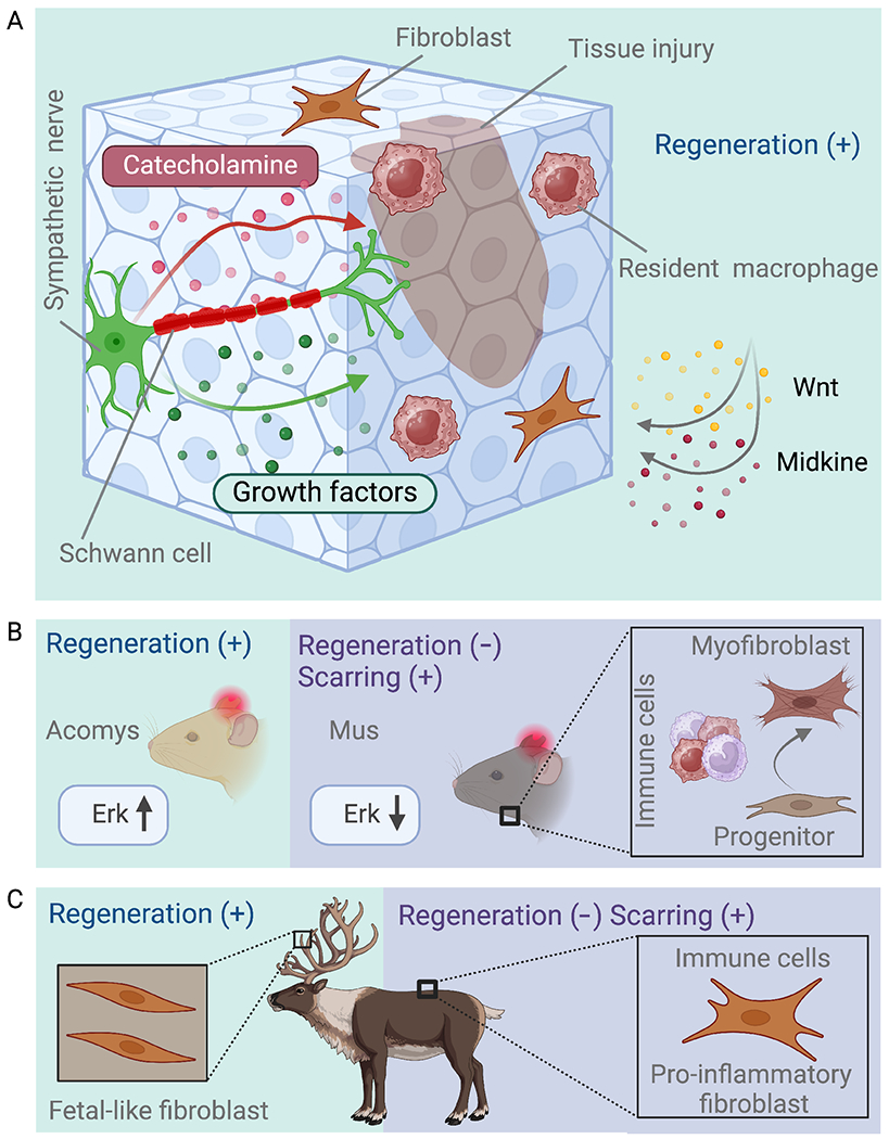
A. For regeneration to supersede scarring, several cell types play a supportive role. Immune cells such as pro-regenerative macrophages, nerve cells and fibroblasts can produce cues for source cells.
B. (Left) Spiny mice, but not house mice, can regenerate ear hole punches due in part to sustained high Erk signaling. (Right) Progenitors located in the mouse dermal fascia generate fibroblasts with pro-scarring, pro-inflammatory profiles that recruit immune cells, while also producing myofibroblasts that promote wound contraction.
C. Antler velvet contains fibroblasts with a pro-regenerative profile that do not recruit immune cells. On the contrary, back skin has pro-scarring fibroblasts with a pro-inflammatory profile.
Immune cells.
Immune cells play a major role in tissue regeneration and exhibit extensive heterogeneity. Prevailing immune cell subpopulations and their kinetics define whether regeneration is favored or inhibited after injury. For example, in zebrafish skeletal muscle, a specific macrophage subset populates injury border zones and secretes the cytokine nicotinamide phosphoribosyltransferase to promote the activity of muscle stem cells121. Rapid installment of anti-inflammatory macrophages and clearance of neutrophils, collagen, and tempering of the fibrotic response have all been associated with sustained regeneration across tissue types and species122,123 124–126. Tissue-resident macrophages, not recruited from bone marrow monocytes, have been shown to be abundant in the context of a pro-regenerative environment, as for instance the injured neonatal mouse heart. In contrast, an influx of non-tissue resident, circulating macrophages predominates in the poorly regenerating adult mouse heart127. Yet, other studies that investigated lung and skin repair, indicate that monocytes/macrophages recruited from the circulation can be pro-regenerative128,129. It is currently unknown whether a unifying principle can be extracted regarding the role of macrophages in diverse tissue regeneration contexts or whether each tissue context shows distinctive interactions with macrophage subsets.
What attracts pro-regenerative macrophages and in which ways do macrophages encourage tissue regeneration? Macrophage involvement in regeneration is one of the more popular areas of study in the field, and only a fraction of available information can be covered here. Genetic evidence from appendage regeneration in zebrafish and axolotl have implicated Wnt and Midkine factors in directing macrophages to sites of regeneration126,130. In mouse skin, macrophage recruitment is mediated by lipids released by wound-adjacent adipocytes131. Evidence in zebrafish indicates that TNF-α secreted by macrophages promotes neurogenesis in radial glial cells through the Tnfrsf1a receptor during spinal cord regeneration in larval zebrafish. In contrast, later-stage macrophages act to decrease levels of IL1B, a cytokine that promotes regeneration132,133. Other innate immune cell types also modulate regeneration, including innate lymphoid cells and neutrophils. For instance, thymic natural killer T cells (NKTs) restrain regeneration of amputated mouse digit tips, whereas splenic NKTs have a pro-regenerative influence134.
Adaptive immunity is increasingly recognized as a contributor to regeneration outcomes. Diverse roles for Treg cells in the regeneration of multiple tissues have emerged after their initial discovery in regenerating skeletal muscle135. These cells have mitogenic roles in regeneration of multiple other tissues including heart, skin, and supporting the intestinal stem cell niche136–138. A comparison of regenerating organs in zebrafish, including retina, spinal cord, and heart, revealed that Treg cells show common and organ-specific expression of regeneration-promoting factors, including Neurotrophin3 in the spinal cord, Neuregulin1 in the heart, and Igf1 in the retina139. Whether Treg cells acquire tissue-specific programs upon injury and regeneration, or instead pre-exist in these tissues is not fully understood. Beyond Treg cells, other types of T cells have also been implicated in local responses to tissue trauma. Wounded skin relies on gamma delta T cells to provide Fgf9 that elicits hair follicle neogenesis in large skin wounds and IL1 to promote hair follicle stem cell proliferation140,141. Furthermore IL-17A production from Th17 cells, a response that is normally associated with protection against microbiota after skin disruption, was recently implicated in the regeneration of sensory nerves142.
Nerves.
Salamander limb regeneration depends on innervation. This observation was initially made in the early 19th century and foreshadowed contemporary findings indicating a contribution of the nervous system in many regeneration contexts143. Early studies of limb regeneration found that the quantity of innervation rather than the nerve type (motor or sensory) was important, and trophic factors were proposed to play a dominant role144. Since then, many proteinaceous “growth factors” that may substitute nerves for blastema formation have been identified, including Neuregulin, FGF2 GDF5, and BMP7, as well as Anterior gradient and transferrin145 146,147 148,149. Many of these factors are thought to act directly on mesenchymal blastema cells to promote their proliferation, although Anterior gradient is proposed to act by relaying nerve signaling to the epidermis150. Denervation of mouse digit tips by sciatic nerve resection causes defects in regeneration, with growth factors PDGF-AA or OSM able to partially rescue these defects. Innervation has also been associated with maintaining high FGF2 levels that promote cell proliferation151,152. Experiments employing digit nerve resection, by contrast, indicate no requirement for regeneration after tip amputation153. In the mouse skin, nerve-derived Shh ligand encourages hair follicle stem cells in the bulge at telogen stage to contribute to anagen stage hair follicles; surprisingly, Shh also shapes a niche that allows upper bulge cells to migrate into the regenerating epidermis154. Such signals also derive from supporting Schwann cells that in the regenerating mouse digit tip can be a source of TGF-B, which initiates skin healing, or OSM and PDGF-AA, which promote blastemal cell proliferation152,155.
Beyond classical growth factors, signaling of sympathetic nerves by catecholamine activation of adrenergic receptors has emerged in several contexts of stem cell activity during regeneration. In mice, ablation of sensory and sympathetic nerves depletes the bone marrow of HSCs, in which B2 and B3 adrenergic signaling from nerves is required to sustain the niche156. These Leptin receptor-positive (Lepr+) stromal niche cells express Nerve growth factor that promotes innervation of the bone marrow, and the nerves in turn activate B adrenergic signaling in the Lepr+ cells to sustain the HSC niche157. Stressful conditions affect hair follicles through increased B adrenergic signaling, driving hyperproliferation, depletion of melanocyte stem cells and hair graying158. Furthermore, channel activity is implicated in the process of innervation during early stages of zebrafish fin regeneration, as revealed by conditional genetic mutants in the major neuronal voltage-gated sodium channel (VGSC) gene scn8ab159.
Cells communicate not only as pairs but within complex circuits of cell-cell communication, choreographing the cell behaviors required from different actors. Recently, a pharmacological screen led to a model in which heart regeneration in zebrafish depends on nerve-derived adrenergic signaling, acting through a subset of macrophages that ultimately influence ECM deposition and cardiomyocyte proliferation160. In another example of cellular coordination, the early interactions of nerves and macrophages with the wound epidermis was proposed to create a niche for blastema cell proliferation and expansion in regenerating axolotl limbs130. These types of multicellular relays are now the focus of combined theoretical modeling and experimental approaches that indicate that the sum of multi-cellular interactions may define regeneration outcomes161,162.
Fibroblasts.
Long considered to be a homogeneous cell type that maintains structural integrity and responds to injury by secreting matrix components, such as collagen, fibroblasts are receiving an increasing amount of attention for their diversity of programs and functions. The classic fibroblast injury response provides a quick fix for major injuries to tissues, such as the skin or the heart. There are obvious benefits to quick-scarring mechanisms that close surface wounds, preserve barrier function, and fill in the gaps of lost myocardium after an ischemic injury. Yet, tissue biomechanics and function are often downgraded after these quick repair mechanisms and the area retains susceptibilities. Animal responses to trauma have thus often been framed as “Regeneration versus Scarring”; and there is truth to this. For instance, zebrafish hearts will heal by scarring if cardiomyogenesis is experimentally blocked, and murine hearts show more effective repair when cardiac fibroblasts are depleted by a CAR-T strategy or a small percentage of them is reprogrammed into myocyte-like cells163–165. Similarly, pericyte-derived fibroblast-like cells contribute to spinal cord injury scars, and genetically disabling division of pericyte progeny in mice improves repair and connectivity after a cord hemisection injury166.
Sequencing experiments have made it clear that fibroblasts constitute a heterogeneous population. Like immune cell types, pro-regenerative and anti-regenerative (pro-scarring) resident fibroblasts can help dictate injury outcomes. In mice, two skin fibroblast types with differential fibrotic injury responses exist that once expressed the transcription factor Engrailed 1 (En1) during embryonic development (EN1 lineage-positive fibroblasts (EPFs) and EN1-naiïve fibroblasts (ENFs))167. EPFs arise at a point in embryonic development in which scarring of skin wounds replaces a more regenerative, scar-free response168, and they are dominant contributors to scarring in adult skin167. ENFs normally adjudicate skin development and fetal regenerative responses to injury168, although a subpopulation of ENFs can induce En1 expression and acquire EPF properties in adult skin wounds169. Perhaps analogously, Erk signaling has been proposed to toggle regenerative and scarring properties of fibroblasts in mice. Spiny mice (Acomys) have an enhanced capacity to regenerate ear hole punches and large back skin wounds as compared to house mice (Mus)170. Their healing responses are associated with sustained high Erk signaling171 (Fig 4B). Erk inhibition promotes scarring in Acomys, whereas sustained Erk activation promotes regeneration in Mus, and differentially Erk-responsive fibroblast subsets are suspected to contribute to species differences171.
Pro-regenerative and pro-scarring fibroblast subtypes display distinct immunomodulatory profiles and properties. Progenitors located in the fascia of mouse dermis undergo a sequence of steps to generate fibroblasts with pro-inflammatory profiles that recruit immune cells, such as macrophages and monocytes. These fibroblasts subsequently convert into myofibroblasts that promote wound contraction172 (Fig 4B). Analogous transitional events happen in injured lungs to generate pro-fibrotic alveolar fibroblasts173. Velvet covering the antlers of reindeer – massive bony appendages cast and regenerated each year – regenerate large hole punches without scarring. Antler velvet contains fibroblasts retaining a fetal profile in contrast to immune-activating profiles of fibroblasts from poorly regenerating back skin. Inflammatory cell recruitment by velvet fibroblasts is muted compared to back skin fibroblasts, even when velvet fibroblasts are grafted ectopically onto back skin followed by injury, indicating that fibroblast-immune cell crosstalk measurably determines regenerative capacity174 (Fig 4C).
4. Size control and tissue integration
Positional, or geometric, memory is a defining feature of regeneration, which brings back what it has lost and only that, weeks to months to years after the initial formation of that lost structure during embryogenesis. These events require mechanisms to retain or instill memory of the correct programs for cell identity in the injured tissue stump, as well as mechanisms to create scale to the correct adult size. As the myriad cellular constituents of tissues return at appropriate proportions, they must be physically and, in some cases, electrically, integrated to regenerate function of that lost organ or appendage.
Epigenetic memory of position.
Trauma unleashes widespread transcriptional changes that ultimately yield to regeneration of order -- cell types return in correct proportion and pattern. This occurs at least in part by recalling epigenetic memories of cell and positional identities that template the pattern and determine the spectrum of cell identities that are produced during regeneration. It is likely that processes to establish and follow epigenetic settings for cell lineage and positional states help regenerating tissues steer clear of cancer programs.
Salamander limb regeneration relies on stably sustained cellular position codes that exist in each limb segment (upper limb, lower limb, hand/foot) as well as anterior-posterior (thumb-pinky) and dorsal-ventral (palm-back of hand)175. Here, fibroblasts maintain stable identities according to proximodistal limb region and radial location around the limb, in this way guiding regeneration of the correct structures. Fibroblasts within different limb segments launch distinct homeodomain expression programs that constrain them to regenerate their own segment and those more distal but not more proximal seqments. This positional constraint arises at least in part from the repressive histone mark H3K27me3 that differentially occupies homeodomain protein genes in fibroblasts from the different limb segments. In the upper arm, the hand-associated Hox gene, such as HoxA13 is occupied by H3K27me3 marks and repressed, so that an upper arm gene like Meis fires early during upper arm regeneration. In contrast, in the hand, HoxA13 has an underoccupied H3K27me3 signature and has corresponding elevated gene expression during hand regeneration 176,177. In essence, limb amputation prompts the resultant blastema to re-initiate the limb development program at the segment level of the amputation.
Radially arranged cell identities are exploited by the limb to help ensure that regeneration occurs only upon amputation and not after simple wounding, by establishing a co-dependent signaling relationship between cells derived from the anterior and posterior limb178. Posterior fibroblasts, as they transform into blastema cells, recreate a key signaling center -- localized Shh expression -- that signals to anterior limb progenitor cells to promote growth and patterning. The epigenetic basis of this memory of posterior limb cells to induce Shh relies on a sustained transcriptional circuit in adult tissue (Fig 5A). Expression of the posterior transcription factor Hand2 persists in the mature limb tissue in axolotl, as well as in mature pectoral fins of zebrafish, which also maintains patterned domains of the anterior transcription factor alx4a and other genes179,180. Hand2 is necessary and sufficient to produce posterior blastema cells that initiate Shh gene expression (Fig 5A). A similar system exists long the D/V axis involving expression of the transcription factor Lmx1b181.
Fig 5. Size control and tissue integration.
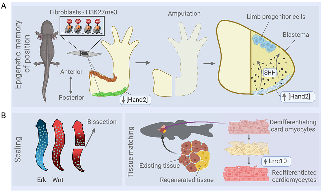
A. Epigenetic memory of position is represented at least in part from the repressive histone mark H3K27me3 in fibroblasts of different limb segments. Posterior fibroblasts of the salamander blastema express the transcription factor Hand2 that initiates SHH signaling to progenitor cells located in the anterior limb.
B. (Left) Wnt and Erk signaling across the body axis of planaria. After animal bissection, the Wnt signaling gradient rapidly re-scales, re-directing neoblasts to form proportionally scaled organs. (Right) Regenerated cardiomyocytes integrate with existing heart muscle in zebrafish, involving maturation and upregulation of Lrrc10 after proliferation.
Positional memory also exists in mice and humans, and transcriptional profiling of fibroblasts across the body has identified positional codes of gene expression that appear to define regions, even after culturing in vitro. Similarly, HOXA11 is specifically expressed in lower arm but not upper arm fibroblasts of adult mice, and lineage and functional studies uncovered a role for this spatially restricted transcription factor in fracture repair 182–184. A current unknown in cross species comparisons is whether mammalian stem cells can reactivate the sequential expression of Hox genes, as observed in regenerating salamander limbs176.
Functional integration of regenerated tissue.
Regeneration outmatches current bioengineered devices in its return of function. For instance, recreating the sensory ability of an arm and its digits is a major challenge for prosthetic device. Tissue matchmaking, to form a seamless interface between old and new tissue, helps regeneration reach its functional goals. For example, if the salamander limb is amputated in the humerus, how does the remaining humerus part connect to its new skeletal elements? Riquelme-Guzmán and colleagues found that continuity between the old bone with regenerated bone relies on osteoclasts to trim the bone at the amputation plane185. During limb regeneration, an intermediate mini-limb is formed and expands to match the size of the animal. Thus, embryonic-like fields of cells have been specified early to make tissues representing limb regions, whereas cells within each tissue compartment continue to proliferate and promote tissue expansion. By denervating limbs at late stages of limb regeneration, a study indicated that nerve input is required for the late, expansion phase of growth186. The authors proposed that the amount of nervous tissue might be a yardstick by which proliferative cells within limb regions hit their target sizes186.
For the heart to effectively regenerate lost muscle, electrical and mechanical coupling of newly formed cardiomyocytes with existing heart tissue is critical. Relevant to this, induced pluripotent stem cell (iPSC)-derived cardiomyocytes that possess fetal morphology and physiology survive (Fig 5B), integrate, and function when introduced into hosts as cell suspensions or patches, yet they may stay immature and can cause arrhythmogenesis187,188. When innate regeneration through dedifferentiation and proliferation of endogenous cardiomyocytes occurs, as in zebrafish, cardiomyocytes at the injury border zone first show embryonic-type physiology as in the nature of calcium ion fluxes189 (Fig 5B). Upregulation of the leucine rich repeat protein (Lrrc10) is required to help cardiomyocytes mature away from the embryonic phenotype (Fig 5B). This maturation occurs by ensuring the structural integrity of the nascent cardiac dyad and modulation of calcium ion dynamics. Lrrc10, first identified in zebrafish heart regeneration190, also induces structural maturation in human iPSC-derived cardiomyocytes189. Thus, cardiomyocyte regeneration requires appropriate redifferentation as well as dedifferentiation189,191, and this knowledge might guide therapeutic applications.
Scaling.
Achieving size during regeneration can occur in multiple ways. Salamander limbs form a blastema that gradually differentiates into the appropriate limb structures, whereas zebrafish fins maintain a blastemal growth zone at their distal tips and add bone segment upon segment until length is achieved. Paradoxically, the crowned king of mammalian regeneration, the liver, reconstitutes an organ-to-body mass ratio from intact lobules after partial hepatectomy, but does not reconstruct the missing lobule from a primordium. Regeneration of liver mass relies at least in part on systemic FGF15 secreted from the gut, that acts through the Klotho receptor, along with liver-derived RSPO that coordinates Wnt and other signaling pathways192,193. At this tissue level, the liver is zonated with hepatocytes expressing different enzymes depending on their distance along the portal-to-central vein axis, the unit structure of the liver. Transcriptome sequencing and genetic fate-mapping has revealed differences in contributions to regeneration from the different zones of hepatocytes, based on these locations with the lobes194–196. This organization is faithfully recapitulated after injury, and depends on LGR4 and LGR5 signaling, whereby the expression of these receptors is stratified along the zones. This signaling helps to control the expansion or contraction of different hepatocyte populations to attain an appropriate proportionality196 193. Interestingly, somatic mutations in growth-promoting genes acquired during development can influence and even increase the regenerative capacity of hepatocyte clones in liver disease197.
Proportional scaling with faithful spatial patterning is a hallmark of whole-body regeneration in planarians. Flatworms grow and shrink according to nutrient availability, and, after bissection, rescale their anatomy although the regenerating bodies are a fraction of their previous size. Sustained positional transcriptional states are important, in which body wall muscle cells display a positional code of signaling molecules, including regulators of Wnt and BMP signaling pathways, that guide neoblasts in building position-appropriate body parts198,199. In planarians, Wnt and ERK signaling are graded across the body axis to control neoblast differentiation and determine the head-to-tail axis and its proportionality200–203. After animal bissection, the Wnt signaling gradient rapidly re-scales, re-directing neoblasts in the severed body to form organs appropriate to the new scaling. Thus, a phoenix-like transformation of body part into whole organism occurs (Fig 5B). How Wnt signaling gradients span millimeter distances is incompletely understood. In particular, whether true gradients of single morphogens exist across such distances or whether these are relay mechanisms between cells is not clear. Dynamic signaling patterns are also represented in regenerating zebrafish scales, in which live imaging of ERK kinase activity revealed a timed series of signaling waves in osteoblasts, traveling millimeter distances in a ringed pattern from the center out to the periphery of the scale204. Each time a wave hits a region of osteoblasts, they see a transient burst of growth. How spatially regulated upstream and downstream players are controlled and sculpt tissue is central to regeneration and relevant to engineering approaches in adult-scale regenerating organs.
Knowing when to stop is important not only when writing literature reviews, but also during regeneration. Several zebrafish mutants that flaunt lengthened fins have shed light on this important issue. Two of the underlying mutations affect the activity of components of potassium leak channels, Kcnk5b and Slc12a7a/Kcc4a, and they cause fins to grow longer with lengthier bony segments, both ontogenetically and during regeneration205,206. The activities of these proteins are linked to signaling through regulation of Kcnk5b phosphorylation by the phosphatase calcineurin, affecting channel conductance207–209. Within this signaling network, another potassium channel, Kcnh2a, acts upstream of calcineurin to control cessation of fin growth210. Further examination of developmental gene expression in channel mutants revealed upregulation of key signaling factors and a tissue phenotype of a larger blastema and faster skeletal growth208. This phenotype is reminiscent of the effects of agents that influence bioelectric signaling and elicit ectopic regenerates in amputated Xenopus limbs211. Together, these observations suggest that cellular ionic setpoints interface with signaling pathways and developmental gene regulatory networks to manage regeneration of entire structures.
Perspectives
Each hallmark mentioned here has knowledge gaps to be filled by discovery research and technological advances. A pipeline of more advanced animal and plant models of regeneration will help identify answers as well as new questions. Although single-cell RNA sequencing has increased the cellular resolution of regenerative events, it does not obviate the need for direct visualization of cellular and subcellular events in live, regenerating tissue. There is an infinite space for the generation of new tools and the combination of these tools with advanced imaging platforms. Importantly, a growing number of studies of developing tissue integrate mechanical cues with molecular signals and cellular behaviors. Mechanical signals are also implicated in regeneration, for example by physically enabling or inhibiting cell division, or by controlling factor release or localization212–214. Advancing methods to spatiotemporally control mechanical influences in injured and regenerating tissues will enable a full appreciation of their impact.
The most common question regenerative biologists receive is “Why?” -- why do certain species and tissues have great regenerative capacity when others are so limited? It is challenging to directly address this topic with rigorously tested hypotheses; one is more likely to accidentally bump into illuminating evidence by exploring how regeneration works. An important arm of trying to address the evolution of regenerative capacity is to understand how enhancers, silencers, insulators or other non-coding DNA elements became associated with protein coding components used earlier in initial embryonic development.
A bright future lies ahead in harvesting the growing body of conceptual and molecular information to engineer regeneration-competent cell states and tissue environments (Fig 6). There are many markers on the road to that destination. Although it is recognized that influential aspects of regenerative biology, such as inflammation and signal presence, can be modulated, the ability to alter intrinsic epigenetic states to access memories of developmental competence is a roadblock that must be overcome215 (Fig. 5). A better understanding of the factors that allow controlled transdifferentiation in certain cell types and contexts, but not in others, will aid in this pursuit. Modulation itself needs precision for safety and efficacy, and this precision can be enforced by incorporating regeneration enhancer elements to dose and target pro-regenerative factors, for instance within gene therapy vectors216. Building synthetic circuits for regeneration based on engineered ligand systems can also infuse elements of control and amplitude. Finally, highly regenerative organisms remind us that stem cell location within a regenerating primordium can impact the competency to express key morphogens and other signaling molecules. Directed spatial targeting of competent stem cells may be most helpful in future applications of regenerative medicine.
Fig 6. Representation of hallmarks of regeneration in proficient and inefficient models, and potential interventions at bottlenecks.

Left: Key cellular and signaling events occur during limb regeneration in the salamander. Middle: Human limb injury leads to fibrosis without restoration of tissue patterns. Right: Manipulation of gene expression states with modern tools such as CRISPR-mediated transcriptional activation (CRISPRa), immunomodulation, and restitution of developmental genetic circuits can help unleash a regenerative response.
Acknowledgments
We apologize to authors for omitting depth or discussion of their field or work due to space constraints. We thank Emmanouella Chatzidaki for artwork and Junsu Kang, Purushothama Tata, and Fei Sun for comments on the manuscript. Figures were created with Biorender.com. KDP acknowledges funding from NIH (R35 HL150713, R01 AR076342, R01 HD115266, R01 HD105033). EMT acknowledges funding from ERC, FWF, and IMP.
Footnotes
Publisher's Disclaimer: This is a PDF file of an unedited manuscript that has been accepted for publication. As a service to our customers we are providing this early version of the manuscript. The manuscript will undergo copyediting, typesetting, and review of the resulting proof before it is published in its final form. Please note that during the production process errors may be discovered which could affect the content, and all legal disclaimers that apply to the journal pertain.
References
- 1.Hsu YC, and Fuchs E (2022). Building and Maintaining the Skin. Cold Spring Harb Perspect Biol 14. 10.1101/cshperspect.a040840. [DOI] [PMC free article] [PubMed] [Google Scholar]
- 2.Hindi SM, and Millay DP (2022). All for One and One for All: Regenerating Skeletal Muscle. Cold Spring Harb Perspect Biol 14. 10.1101/cshperspect.a040824. [DOI] [PMC free article] [PubMed] [Google Scholar]
- 3.Biermann M, and Reya T (2022). Hematopoietic Stem Cells and Regeneration. Cold Spring Harb Perspect Biol 14. 10.1101/cshperspect.a040774. [DOI] [PMC free article] [PubMed] [Google Scholar]
- 4.Wagner DE, Wang IE, and Reddien PW (2011). Clonogenic neoblasts are pluripotent adult stem cells that underlie planarian regeneration. Science 332, 811–816. 10.1126/science.1203983. [DOI] [PMC free article] [PubMed] [Google Scholar]
- 5.Zeng A, Li H, Guo L, Gao X, McKinney S, Wang Y, Yu Z, Park J, Semerad C, Ross E, et al. (2018). Prospectively Isolated Tetraspanin(+) Neoblasts Are Adult Pluripotent Stem Cells Underlying Planaria Regeneration. Cell 173, 1593–1608 e1520. 10.1016/j.cell.2018.05.006. [DOI] [PMC free article] [PubMed] [Google Scholar]
- 6.Raz AA, Wurtzel O, and Reddien PW (2021). Planarian stem cells specify fate yet retain potency during the cell cycle. Cell Stem Cell 28, 1307–1322 e1305. 10.1016/j.stem.2021.03.021. [DOI] [PMC free article] [PubMed] [Google Scholar]
- 7.Reddien PW (2022). Positional Information and Stem Cells Combine to Result in Planarian Regeneration. Cold Spring Harb Perspect Biol 14. 10.1101/cshperspect.a040717. [DOI] [PMC free article] [PubMed] [Google Scholar]
- 8.Ricci L, and Srivastava M (2021). Transgenesis in the acoel worm Hofstenia miamia. Dev Cell 56, 3160–3170 e3164. 10.1016/j.devcel.2021.10.012. [DOI] [PMC free article] [PubMed] [Google Scholar]
- 9.Poss KD, Wilson LG, and Keating MT (2002). Heart regeneration in zebrafish. Science 298, 2188–2190. [DOI] [PubMed] [Google Scholar]
- 10.Kikuchi K, Holdway JE, Werdich AA, Anderson RM, Fang Y, Egnaczyk GF, Evans T, Macrae CA, Stainier DY, and Poss KD (2010). Primary contribution to zebrafish heart regeneration by gata4(+) cardiomyocytes. Nature 464, 601–605. nature08804 [pii] 10.1038/nature08804. [DOI] [PMC free article] [PubMed] [Google Scholar]
- 11.Honkoop H, de Bakker DE, Aharonov A, Kruse F, Shakked A, Nguyen PD, de Heus C, Garric L, Muraro MJ, Shoffner A, et al. (2019). Single-cell analysis uncovers that metabolic reprogramming by ErbB2 signaling is essential for cardiomyocyte proliferation in the regenerating heart. Elife 8. 10.7554/eLife.50163. [DOI] [PMC free article] [PubMed] [Google Scholar]
- 12.Zhu W, Zhang E, Zhao M, Chong Z, Fan C, Tang Y, Hunter JD, Borovjagin AV, Walcott GP, Chen JY, et al. (2018). Regenerative Potential of Neonatal Porcine Hearts. Circulation 138, 2809–2816. 10.1161/CIRCULATIONAHA.118.034886. [DOI] [PMC free article] [PubMed] [Google Scholar]
- 13.Ye L, D’Agostino G, Loo SJ, Wang CX, Su LP, Tan SH, Tee GZ, Pua CJ, Pena EM, Cheng RB, et al. (2018). Early Regenerative Capacity in the Porcine Heart. Circulation 138, 2798–2808. 10.1161/CIRCULATIONAHA.117.031542. [DOI] [PubMed] [Google Scholar]
- 14.Porrello ER, Mahmoud AI, Simpson E, Hill JA, Richardson JA, Olson EN, and Sadek HA (2011). Transient regenerative potential of the neonatal mouse heart. Science 331, 1078–1080. 331/6020/1078 [pii] 10.1126/science.1200708. [DOI] [PMC free article] [PubMed] [Google Scholar]
- 15.Li X, Wu F, Gunther S, Looso M, Kuenne C, Zhang T, Wiesnet M, Klatt S, Zukunft S, Fleming I, et al. (2023). Inhibition of fatty acid oxidation enables heart regeneration in adult mice. Nature 622, 619–626. 10.1038/s41586-023-06585-5. [DOI] [PMC free article] [PubMed] [Google Scholar]
- 16.Puente BN, Kimura W, Muralidhar SA, Moon J, Amatruda JF, Phelps KL, Grinsfelder D, Rothermel BA, Chen R, Garcia JA, et al. (2014). The Oxygen-Rich Postnatal Environment Induces Cardiomyocyte Cell-Cycle Arrest through DNA Damage Response. Cell 157, 565–579. 10.1016/j.cell.2014.03.032. [DOI] [PMC free article] [PubMed] [Google Scholar]
- 17.Tanaka HV, Ng NCY, Yang Yu Z, Casco-Robles MM, Maruo F,Tsonis PA, and Chiba C (2016). A developmentally regulated switch from stem cells to dedifferentiation for limb muscle regeneration in newts. Nat Commun 7, 11069. 10.1038/ncomms11069. [DOI] [PMC free article] [PubMed] [Google Scholar]
- 18.Sandoval-Guzman T, Wang H, Khattak S, Schuez M, Roensch K, Nacu E, Tazaki A, Joven A, Tanaka EM, and Simon A (2014). Fundamental differences in dedifferentiation and stem cell recruitment during skeletal muscle regeneration in two salamander species. Cell Stem Cell 14, 174–187. 10.1016/j.stem.2013.11.007. [DOI] [PubMed] [Google Scholar]
- 19.Konkimalla A, Tata A, and Tata PR. (2022). Lung Regeneration: Cells, Models, and Mechanisms. Cold Spring Harb Perspect Biol 14. 10.1101/cshperspect.a040873. [DOI] [PMC free article] [PubMed] [Google Scholar]
- 20.Tata PR, Mou H, Pardo-Saganta A, Zhao R, Prabhu M, Law BM, Vinarsky V, Cho JL, Breton S, Sahay A, et al. (2013). Dedifferentiation of committed epithelial cells into stem cells in vivo. Nature 503, 218–223. 10.1038/nature12777. [DOI] [PMC free article] [PubMed] [Google Scholar]
- 21.Chen Y, Luttmann FF, Schoger E, Scholer HR, Zelarayan LC, Kim KP, Haigh JJ, Kim J, and Braun T (2021). Reversible reprogramming of cardiomyocytes to a fetal state drives heart regeneration in mice. Science 373, 1537–1540. 10.1126/science.abg5159. [DOI] [PubMed] [Google Scholar]
- 22.Shelby H, Shelby T, and Wernig M (2022). Somatic Lineage Reprogramming. Cold Spring Harb Perspect Biol 14. 10.1101/cshperspect.a040808. [DOI] [PMC free article] [PubMed] [Google Scholar]
- 23.Hishida T, Yamamoto M, Hishida-Nozaki Y, Shao C, Huang L, Wang C, Shojima K, Xue Y, Hang Y, Shokhirev M, et al. (2022). In vivo partial cellular reprogramming enhances liver plasticity and regeneration. Cell Rep 39, 110730. 10.1016/j.celrep.2022.110730. [DOI] [PMC free article] [PubMed] [Google Scholar]
- 24.Wang C, Rabadan Ros R, Martinez-Redondo P, Ma Z, Shi L, Xue Y, Guillen-Guillen I, Huang L, Hishida T, Liao HK, et al. (2021). In vivo partial reprogramming of myofibers promotes muscle regeneration by remodeling the stem cell niche. Nat Commun 12, 3094. 10.1038/S41467-021-23353-z. [DOI] [PMC free article] [PubMed] [Google Scholar]
- 25.Monroe TO, Hill MC, Morikawa Y, Leach JP, Heallen T, Cao S, Krijger PHL, de Laat W, Wehrens XHT, Rodney GG, and Martin JF (2019). YAP Partially Reprograms Chromatin Accessibility to Directly Induce Adult Cardiogenesis In Vivo. Dev Cell 48, 765–779 e767. 10.1016/j.devcel.2019.01.017. [DOI] [PMC free article] [PubMed] [Google Scholar]
- 26.Pajcini KV, Corbel SY, Sage J, Pomerantz JH, and Blau HM (2010). Transient inactivation of Rb and ARF yields regenerative cells from postmitotic mammalian muscle. Cell Stem Cell 7, 198–213. 10.1016/j.stem.2010.05.022. [DOI] [PMC free article] [PubMed] [Google Scholar]
- 27.Vergara MN, Tsissios G, and Del Rio-Tsonis K (2018). Lens regeneration: a historical perspective. Int J Dev Biol 62, 351–361. 10.1387/ijdb.180084nv. [DOI] [PMC free article] [PubMed] [Google Scholar]
- 28.Eguchi G, Abe SI, and Watanabe K (1974). Differentiation of lens-like structures from newt iris epithelial cells in vitro. Proc Natl Acad Sci U S A 71, 5052–5056. 10.1073/pnas.71.12.5052. [DOI] [PMC free article] [PubMed] [Google Scholar]
- 29.Suetsugu-Maki R, Maki N, Nakamura K, Sumanas S, Zhu J, Del Rio-Tsonis K, and Tsonis PA (2012). Lens regeneration in axolotl: new evidence of developmental plasticity. BMC Biol 10, 103. 10.1186/1741-7007-10-103. [DOI] [PMC free article] [PubMed] [Google Scholar]
- 30.Freeman G. (1963). Lens Regeneration from the Cornea in Xenopus Laevis. J Exp Zool 154, 39–65. 10.1002/jez.1401540105. [DOI] [PubMed] [Google Scholar]
- 31.Todd L, and Reh TA (2022). Comparative Biology of Vertebrate Retinal Regeneration: Restoration of Vision through Cellular Reprogramming. Cold Spring Harb Perspect Biol 14. 10.1101/cshperspect.a040816. [DOI] [PMC free article] [PubMed] [Google Scholar]
- 32.Del Rio-Tsonis K, and Tsonis PA (2003). Eye regeneration at the molecular age. Dev Dyn 226, 211–224. 10.1002/dvdy.10224. [DOI] [PubMed] [Google Scholar]
- 33.Fausett BV, and Goldman D (2006). A role for alpha1 tubulin-expressing Muller glia in regeneration of the injured zebrafish retina. J Neurosci 26, 6303–6313. 10.1523/JNEUROSCI.0332-06.2006. [DOI] [PMC free article] [PubMed] [Google Scholar]
- 34.Thorel E, Nepote V, Avril I, Kohno K, Desgraz R, Chera S, and Herrera PL. (2010). Conversion of adult pancreatic alpha-cells to beta-cells after extreme beta-cell loss. Nature 464, 1149–1154. 10.1038/nature08894. [DOI] [PMC free article] [PubMed] [Google Scholar]
- 35.Goldenring JR, and Mills JC (2022). Cellular Plasticity, Reprogramming, and Regeneration: Metaplasia in the Stomach and Beyond. Gastroenterology 162, 415–430. 10.1053/j.gastro.2021.10.036. [DOI] [PMC free article] [PubMed] [Google Scholar]
- 36.Liang R, Lin YH, and Zhu H (2021). Genetic and Cellular Contributions to Liver Regeneration. Cold Spring Harb Perspect Biol 14. 10.1101/cshperspect.a040832. [DOI] [PMC free article] [PubMed] [Google Scholar]
- 37.Cigliola V, Ghila L, Thorel F, van Gurp L, Baronnier D, Oropeza D, Gupta S, Miyatsuka T, Kaneto H, Magnuson MA, et al. (2018). Pancreatic islet-autonomous insulin and smoothened-mediated signalling modulate identity changes of glucagon(+) alpha-cells. Nat Cell Biol 20, 1267–1277. 10.1038/s41556-018-0216-y. [DOI] [PMC free article] [PubMed] [Google Scholar]
- 38.Chera S, Baronnier D, Ghila L, Cigliola V, Jensen JN, Gu G, Furuyama K, Thorel F, Gribble FM, Reimann F, and Herrera PL. (2014). Diabetes recovery by age-dependent conversion of pancreatic delta-cells into insulin producers. Nature 514, 503–507. 10.1038/nature13633. [DOI] [PMC free article] [PubMed] [Google Scholar]
- 39.Miyaoka Y, Ebato K, Kato H, Arakawa S, Shimizu S, and Miyajima A (2012). Hypertrophy and unconventional cell division of hepatocytes underlie liver regeneration. Curr Biol 22, 1166–1175. 10.1016/j.cub.2012.05.016. [DOI] [PubMed] [Google Scholar]
- 40.Schaub JR, Huppert KA, Kurial SNT, Hsu BY, Cast AE, Donnelly B, Karns RA, Chen R, Rezvani M, Luu HY, et al. (2018). De novo formation of the biliary system by TGFbeta-mediated hepatocyte transdifferentiation. Nature 557, 247–251. 10.1038/s41586-018-0075-5. [DOI] [PMC free article] [PubMed] [Google Scholar]
- 41.Tarlow BD, Pelz C, Naugler WE, Wakefield L, Wilson EM, Finegold MJ, and Grompe M (2014). Bipotential adult liver progenitors are derived from chronically injured mature hepatocytes. Cell Stem Cell 15, 605–618. 10.1016/j.stem.2014.09.008. [DOI] [PMC free article] [PubMed] [Google Scholar]
- 42.Russell JO, Lu WY, Okabe H, Abrams M, Oertel M, Poddar M, Singh S, Forbes SJ, and Monga SP (2019). Hepatocyte-Specific beta-Catenin Deletion During Severe Liver Injury Provokes Cholangiocytes to Differentiate Into Hepatocytes. Hepatology 69, 742–759. 10.1002/hep.30270. [DOI] [PMC free article] [PubMed] [Google Scholar]
- 43.Raven A, Lu WY, Man TY, Ferreira-Gonzalez S, O’Duibhir E, Dwyer BJ, Thomson JP, Meehan RR, Bogorad R, Koteliansky V, et al. (2017). Cholangiocytes act as facultative liver stem cells during impaired hepatocyte regeneration. Nature 547, 350–354. 10.1038/nature23015. [DOI] [PMC free article] [PubMed] [Google Scholar]
- 44.Leikina E, Gamage DG, Prasad V, Goykhberg J, Crowe M, Diao J, Kozlov MM, Chernomordik LV, and Millay DP (2018). Myomaker and Myomerger Work Independently to Control Distinct Steps of Membrane Remodeling during Myoblast Fusion. Dev Cell 46, 767–780 e767. 10.1016/j.devcel.2018.08.006. [DOI] [PMC free article] [PubMed] [Google Scholar]
- 45.Millay DP, Sutherland LB, Bassel-Duby R, and Olson EN (2014). Myomaker is essential for muscle regeneration. Genes Dev 28, 1641–1646. 10.1101/gad.247205.114. [DOI] [PMC free article] [PubMed] [Google Scholar]
- 46.Lin YH, Zhang S, Zhu M, Lu T, Chen K, Wen Z, Wang S, Xiao G, Luo D, Jia Y, et al. (2020). Mice With Increased Numbers of Polyploid Hepatocytes Maintain Regenerative Capacity But Develop Fewer Hepatocellular Carcinomas Following Chronic Liver Injury. Gastroenterology 158, 1698–1712 e1614. 10.1053/j.gastro.2020.01.026. [DOI] [PMC free article] [PubMed] [Google Scholar]
- 47.Engel FB, Schebesta M, Duong MT, Lu G, Ren S, Madwed JB, Jiang H, Wang Y, and Keating MT (2005). p38 MAP kinase inhibition enables proliferation of adult mammalian cardiomyocytes. Genes Dev 19, 1175–1187. [DOI] [PMC free article] [PubMed] [Google Scholar]
- 48.Soonpaa MH, and Field LJ (1997). Assessment of cardiomyocyte DNA synthesis in normal and injured adult mouse hearts. The American journal of physiology 272, H220–226. [DOI] [PubMed] [Google Scholar]
- 49.Soonpaa MH, Kim KK, Pajak L, Franklin M, and Field LJ (1996). Cardiomyocyte DNA synthesis and binucleation during murine development. The American journal of physiology 271, H2183–2189. [DOI] [PubMed] [Google Scholar]
- 50.Hirose K, Payumo AY, Cutie S, Hoang A, Zhang H, Guyot R, Lunn D, Bigley RB, Yu H, Wang J, et al. (2019). Evidence for hormonal control of heart regenerative capacity during endothermy acquisition. Science 364, 184–188. 10.1126/science.aar2038. [DOI] [PMC free article] [PubMed] [Google Scholar]
- 51.Patterson M, Barske L, Van Handel B, Rau CD, Gan P, Sharma A, Parikh S, Denholtz M, Huang Y, Yamaguchi Y, et al. (2017). Frequency of mononuclear diploid cardiomyocytes underlies natural variation in heart regeneration. Nat Genet 49, 1346–1353. 10.1038/ng.3929. [DOI] [PMC free article] [PubMed] [Google Scholar]
- 52.Gonzalez-Rosa JM, Burns CE, and Burns CG (2017). Zebrafish heart regeneration: 15 years of discoveries. Regeneration (Oxf) 4, 105–123. 10.1002/reg2.83. [DOI] [PMC free article] [PubMed] [Google Scholar]
- 53.Losick VP, Fox DT, and Spradling AC (2013). Polyploidization and cell fusion contribute to wound healing in the adult Drosophila epithelium. Curr Biol 23, 2224–2232. 10.1016/j.cub.2013.09.029. [DOI] [PMC free article] [PubMed] [Google Scholar]
- 54.Cohen E, Allen SR, Sawyer JK, and Fox DT (2018). Fizzy-Related dictates A cell cycle switch during organ repair and tissue growth responses in the Drosophila hindgut. Elife 7. 10.7554/eLife.38327. [DOI] [PMC free article] [PubMed] [Google Scholar]
- 55.Fox DT, and Spradling AC (2009). The Drosophila hindgut lacks constitutively active adult stem cells but proliferates in response to tissue damage. Cell Stem Cell 5, 290–297. 10.1016/j.stem.2009.06.003. [DOI] [PMC free article] [PubMed] [Google Scholar]
- 56.Cigliola V, Shoffner A, Lee N, Ou J, Gonzalez TJ, Hoque J, Becker CJ, Han Y, Shen G, Faw TD, et al. (2023). Spinal cord repair is modulated by the neurogenic factor Hb-egf under direction of a regeneration-associated enhancer. Nat Commun 14, 4857. 10.1038/S41467-023-40486-5. [DOI] [PMC free article] [PubMed] [Google Scholar]
- 57.Goldman JA, Kuzu G, Lee N, Karasik J, Gemberling M, Foglia MJ, Karra R, Dickson AL, Sun F, Tolstorukov MY, and Poss KD (2017). Resolving Heart Regeneration by Replacement Histone Profiling. Dev Cell 40, 392–404 e395. 10.1016/j.devcel.2017.01.013. [DOI] [PMC free article] [PubMed] [Google Scholar]
- 58.Vizcaya-Molina E, Klein CC, Serras F, Mishra RK, Guigo R, and Corominas M (2018). Damage-responsive elements in Drosophila regeneration. Genome Res 28, 1852–1866. 10.1101/gr.233098.117. [DOI] [PMC free article] [PubMed] [Google Scholar]
- 59.Wang W, Hu CK, Zeng A, Alegre D, Hu D, Gotting K, Ortega Granillo A, Wang Y, Robb S, Schnittker R, et al. (2020). Changes in regeneration-responsive enhancers shape regenerative capacities in vertebrates. Science 369. 10.1126/science.aaz3090. [DOI] [PMC free article] [PubMed] [Google Scholar]
- 60.Lee HJ, Hou Y, Chen Y, Dailey ZZ, Riddihough A, Jang HS, Wang T, and Johnson SL (2020). Regenerating zebrafish fin epigenome is characterized by stable lineage-specific DNA methylation and dynamic chromatin accessibility. Genome Biol 21, 52. 10.1186/s13059-020-1948-0. [DOI] [PMC free article] [PubMed] [Google Scholar]
- 61.Suzuki N, Hirano K, Ogino H, and Ochi H (2019). Arid3a regulates nephric tubule regeneration via evolutionarily conserved regeneration signal-response enhancers. Elife 8. 10.7554/eLife.43186. [DOI] [PMC free article] [PubMed] [Google Scholar]
- 62.Gehrke AR, Neverett E, Luo YJ, Brandt A, Ricci L, Hulett RE, Gompers A, Ruby JG, Rokhsar DS, Reddien PW., and Srivastava M. (2019). Acoel genome reveals the regulatory landscape of whole-body regeneration. Science 363. 10.1126/science.aau6173. [DOI] [PubMed] [Google Scholar]
- 63.Jimenez E, Slevin CC, Song W, Chen Z, Frederickson SC, Gildea D, Wu W, Elkahloun AG, Ovcharenko I, and Burgess SM (2022). A regulatory network of Sox and Six transcription factors initiate a cell fate transformation during hearing regeneration in adult zebrafish. Cell Genom 2. 10.1016/j.xgen.2022.100170. [DOI] [PMC free article] [PubMed] [Google Scholar]
- 64.Li L, Cui L, Lin P, Liu Z, Bao S, Ma X, Nan H, Zhu W, Cen J, Mao Y, et al. (2023). Kupffer-cell-derived IL-6 is repurposed for hepatocyte dedifferentiation via activating progenitor genes from injury-specific enhancers. Cell Stem Cell 30, 283–299 e289. 10.1016/j.stem.2023.01.009. [DOI] [PubMed] [Google Scholar]
- 65.Arbach HE, Harland-Dunaway M, Braden C, Chitsazan AD, Pickering E, Patel JH, and Wills AE (2022). Chromatin accessibility analysis reveals distinct functions for HDAC and EZH2 activities in early appendage regeneration. Wound Repair Regen 30, 707–725. 10.1111/wrr.13057. [DOI] [PubMed] [Google Scholar]
- 66.Jia Y, Li L, Lin YH, Gopal P, Shen S, Zhou K, Yu X, Sharma T, Zhang Y, Siegwart DJ, et al. (2022). In vivo CRISPR screening identifies BAZ2 chromatin remodelers as druggable regulators of mammalian liver regeneration. Cell Stem Cell 29, 372–385 e378. 10.1016/j.stem.2022.01.001. [DOI] [PMC free article] [PubMed] [Google Scholar]
- 67.Kang J, Hu J, Karra R, Dickson AL, Tornini VA, Nachtrab G, Gemberling M, Goldman JA, Black BL, and Poss KD (2016). Modulation of tissue repair by regeneration enhancer elements. Nature 532, 201–206. 10.1038/nature17644. [DOI] [PMC free article] [PubMed] [Google Scholar]
- 68.Harris RE, Setiawan L, Saul J, and Hariharan IK (2016). Localized epigenetic silencing of a damage-activated WNT enhancer limits regeneration in mature Drosophila imaginal discs. Elife 5. 10.7554/eLife.11588. [DOI] [PMC free article] [PubMed] [Google Scholar]
- 69.Sun F, Ou J, Shoffner AR, Luan Y, Yang H, Song L, Safi A, Cao J, Yue F, Crawford GE, and Poss KD (2022). Enhancer selection dictates gene expression responses in remote organs during tissue regeneration. Nat Cell Biol 24, 685–696. 10.1038/s41556-022-00906-y. [DOI] [PMC free article] [PubMed] [Google Scholar]
- 70.Zlatanova I, Sun F, Wu RS, Chen X, Lau BH, Colombier P, Sinha T, Celona B, Xu SM, Materna SC, et al. (2023). An injury-responsive mmp14b enhancer is required for heart regeneration. Sci Adv 9, eadh5313. 10.1126/sciadv.adh5313. [DOI] [PMC free article] [PubMed] [Google Scholar]
- 71.Harris RE, Stinchfield MJ, Nystrom SL, McKay DJ, and Hariharan IK (2020). Damage-responsive, maturity-silenced enhancers regulate multiple genes that direct regeneration in Drosophila. Elife 9. 10.7554/eLife.58305. [DOI] [PMC free article] [PubMed] [Google Scholar]
- 72.Begeman IJ, Shin K, Osorio-Mendez D, Kurth A, Lee N, Chamberlain TJ, Pelegri FJ, and Kang J (2020). Decoding an organ regeneration switch by dissecting cardiac regeneration enhancers. Development 147. 10.1242/dev.194019. [DOI] [PMC free article] [PubMed] [Google Scholar]
- 73.Ando K, Ou J, Thompson JD, Welsby J, Bangru S, Shen J, Wei X, Diao Y, and Poss KD (2024). A screen for regeneration-associated silencer regulatory elements in zebrafish. Dev Cell. 10.1016/j.devcel.2024.01.004. [DOI] [PMC free article] [PubMed] [Google Scholar]
- 74.Goldman JA, and Poss KD (2020). Gene regulatory programmes of tissue regeneration. Nat Rev Genet 21, 511–525. 10.1038/s41576-020-0239-7. [DOI] [PMC free article] [PubMed] [Google Scholar]
- 75.Sikes JM, and Newmark PA (2013). Restoration of anterior regeneration in a planarian with limited regenerative ability. Nature 500, 77–80. 10.1038/nature12403. [DOI] [PMC free article] [PubMed] [Google Scholar]
- 76.Liu SY, Selck C, Friedrich B, Lutz R, Vila-Farre M, Dahl A, Brandi H, Lakshmanaperumal N, Henry I, and Rink JC (2013). Reactivating head regrowth in a regeneration-deficient planarian species. Nature 500, 81–84. 10.1038/nature12414. [DOI] [PubMed] [Google Scholar]
- 77.Mack KL, Talbott HE, Griffin MF, Parker JBL, Guardino NJ, Spielman AF, Davitt MF, Mascharak S, Downer M, Morgan A, et al. (2023). Allele-specific expression reveals genetic drivers of tissue regeneration in mice. Cell Stem Cell 30, 1368–1381 e1366. 10.1016/j.stem.2023.08.010. [DOI] [PMC free article] [PubMed] [Google Scholar]
- 78.Perez-Garijo A, Shlevkov E, and Morata G (2009). The role of Dpp and Wg in compensatory proliferation and in the formation of hyperplastic overgrowths caused by apoptotic cells in the Drosophila wing disc. Development 136, 1169–1177. 10.1242/dev.034017. [DOI] [PubMed] [Google Scholar]
- 79.Chera S, Ghila L, Dobretz K, Wenger Y, Bauer C, Buzgariu W, Martinou JC, and Galliot B (2009). Apoptotic cells provide an unexpected source of Wnt3 signaling to drive hydra head regeneration. Dev Cell 17, 279–289. 10.1016/j.devcel.2009.07.014. [DOI] [PubMed] [Google Scholar]
- 80.Tseng AS, Adams DS, Qiu D, Koustubhan P, and Levin M (2007). Apoptosis is required during early stages of tail regeneration in Xenopus laevis. Dev Biol 301, 62–69. 10.1016/j.ydbio.2006.10.048. [DOI] [PMC free article] [PubMed] [Google Scholar]
- 81.Coppe JP, Desprez PY, Krtolica A, and Campisi J (2010). The senescence-associated secretory phenotype: the dark side of tumor suppression. Annu Rev Pathol 5, 99–118. 10.1146/annurev-pathol-121808-102144. [DOI] [PMC free article] [PubMed] [Google Scholar]
- 82.Ritschka B, Storer M, Mas A, Heinzmann F, Ortells MC, Morton JP, Sansom OJ, Zender L, and Keyes WM (2017). The senescence-associated secretory phenotype induces cellular plasticity and tissue regeneration. Genes Dev 31, 172–183. 10.1101/gad.290635.116. [DOI] [PMC free article] [PubMed] [Google Scholar]
- 83.Mosteiro L, Pantoja C, Alcazar N, Marion RM, Chondronasiou D, Rovira M, Fernandez-Marcos PJ, Munoz-Martin M, Blanco-Aparicio C, Pastor J, et al. (2016). Tissue damage and senescence provide critical signals for cellular reprogramming in vivo. Science 354. 10.1126/science.aaf4445. [DOI] [PubMed] [Google Scholar]
- 84.Salinas-Saavedra M, Febrimarsa, Krasovec G, Horkan HR, Baxevanis, and Frank U (2023). Senescence-induced cellular reprogramming drives cnidarian whole-body regeneration. Cell Rep 42, 112687. 10.1016/j.celrep.2023.112687. [DOI] [PMC free article] [PubMed] [Google Scholar]
- 85.Yun MH, Davaapil H, and Brockes JP (2015). Recurrent turnover of senescent cells during regeneration of a complex structure. Elife 4. 10.7554/eLife.05505. [DOI] [PMC free article] [PubMed] [Google Scholar]
- 86.Yu Q, Walters HE, Pasquini G, Pal Singh S, Lachnit M, Oliveira CR, Leon-Perinan D , Petzold A, Kesavan P, Subiran Adrados C, et al. (2023). Cellular senescence promotes progenitor cell expansion during axolotl limb regeneration. Dev Cell 58, 2416–2427 e2417. 10.1016/j.devcel.2023.09.009. [DOI] [PubMed] [Google Scholar]
- 87.Paramos-de-Carvalho D, Martins I, Cristovao AM, Dias AF, Neves-Silva D, Pereira T, Chapela D, Farinho A, Jacinto A, and Saude L (2021). Targeting senescent cells improves functional recovery after spinal cord injury. Cell Rep 36, 109334. 10.1016/j.celrep.2021.109334. [DOI] [PubMed] [Google Scholar]
- 88.Kragl M, Knapp D, Nacu E, Khattak S, Maden M, Epperlein HH, and Tanaka EM (2009). Cells keep a memory of their tissue origin during axolotl limb regeneration. Nature 460, 60–65. nature08152 [pii] 10.1038/nature08152. [DOI] [PubMed] [Google Scholar]
- 89.Rinkevich Y, Lindau P, Ueno H, Longaker MT, and Weissman IL (2011). Germ-layer and lineage-restricted stem/progenitors regenerate the mouse digit tip. Nature 476, 409–413. 10.1038/nature10346. [DOI] [PMC free article] [PubMed] [Google Scholar]
- 90.Murata K, Jadhav U, Madha S, van Es J, Dean J, Cavazza A, Wucherpfennig K, Michor F, Clevers H, and Shivdasani RA (2020). Ascl2-Dependent Cell Dedifferentiation Drives Regeneration of Ablated Intestinal Stem Cells. Cell Stem Cell 26, 377–390 e376. 10.1016/j.stem.2019.12.011. [DOI] [PMC free article] [PubMed] [Google Scholar]
- 91.Jadhav U, Saxena M, O’Neill NK, Saadatpour A, Yuan GC, Herbert Z, Murata K, and Shivdasani RA (2017). Dynamic Reorganization of Chromatin Accessibility Signatures during Dedifferentiation of Secretory Precursors into Lgr5+ Intestinal Stem Cells. Cell Stem Cell 21, 65–77 e65. 10.1016/j.stem.2017.05.001. [DOI] [PMC free article] [PubMed] [Google Scholar]
- 92.Jadhav U, Manieri E, Nalapareddy K, Madha S, Chakrabarti S, Wucherpfennig K, Barefoot M, and Shivdasani RA (2020). Replicational Dilution of H3K27me3 in Mammalian Cells and the Role of Poised Promoters. Mol Cell 78, 141–151 e145. 10.1016/j.molcel.2020.01.017. [DOI] [PMC free article] [PubMed] [Google Scholar]
- 93.Naik S, Larsen SB, Gomez NC, Alaverdyan K, Sendoel A, Yuan S, Polak L, Kulukian A, Chai S, and Fuchs E (2017). Inflammatory memory sensitizes skin epithelial stem cells to tissue damage. Nature 550, 475–480. 10.1038/nature24271. [DOI] [PMC free article] [PubMed] [Google Scholar]
- 94.Larsen SB, Cowley CJ, Sajjath SM, Barrows D, Yang Y, Carroll TS, and Fuchs E (2021). Establishment, maintenance, and recall of inflammatory memory. Cell Stem Cell 28, 1758–1774 e1758. 10.1016/j.stem.2021.07.001. [DOI] [PMC free article] [PubMed] [Google Scholar]
- 95.Gonzales KAU, Polak L, Matos I, Tierney MT, Gola A, Wong E, Infarinato NR, Nikolova M, Luo S, Liu S, et al. (2021). Stem cells expand potency and alter tissue fitness by accumulating diverse epigenetic memories. Science 374, eabh2444. 10.1126/science.abh2444. [DOI] [PMC free article] [PubMed] [Google Scholar]
- 96.Levra Levron C, Watanabe M, Proserpio V, Piacenti G, Lauria A, Kaltenbach S, Tamburrini A, Nohara T, Anselmi E, Duval C, et al. (2023). Tissue memory relies on stem cell priming in distal undamaged areas. Nat Cell Biol 25, 740–753. 10.1038/s41556-023-01120-0. [DOI] [PMC free article] [PubMed] [Google Scholar]
- 97.Sun F, and Poss KD (2023). Inter-organ communication during tissue regeneration. Development 150. 10.1242/dev.202166. [DOI] [PMC free article] [PubMed] [Google Scholar]
- 98.Richardson D. (1945). Thyroid and pituitary hormones in relation to regeneration; regeneration of the hind leg of the newt, Triturus viridescens, with different combinations of thyroid and pituitary hormones. J Exp Zool 100, 417–429. 10.1002/jez.1401000308. [DOI] [PubMed] [Google Scholar]
- 99.Wilkerson JA (1963). The Role of Growth Hormone in Regeneration of the Forelimb of the Hypophysectomized Newt. J Exp Zool 154, 223–230. 10.1002/jez.1401540209. [DOI] [PubMed] [Google Scholar]
- 100.Liversage RA, and Scadding SR (1969). Re-establishment of forelimb regeneration in adult hypophysectomized Diemictylus (Triturus) viridescens given frog anterior pituitary extract. J Exp Zool 170, 381–395. 10.1002/jez.1401700402. [DOI] [PubMed] [Google Scholar]
- 101.Zhang M, Chen Y, Xu H, Yang L, Yuan F, Li L, Xu Y, Chen Y, Zhang C, and Lin G (2018). Melanocortin Receptor 4 Signaling Regulates Vertebrate Limb Regeneration. Dev Cell 46, 397–409 e395. 10.1016/j.devcel.2018.07.021. [DOI] [PMC free article] [PubMed] [Google Scholar]
- 102.Grobstein C. (1947). The role of androgen in declining regenerative capacity during morphogenesis of the Platypoecilus maculatus gonopodium. J Exp Zool 106, 313–344. 10.1002/jez.1401060307. [DOI] [PubMed] [Google Scholar]
- 103.Nachtrab G, Czerwinski M, and Poss KD (2011). Sexually dimorphic fin regeneration in zebrafish controlled by androgen/GSK3 signaling. Curr Biol 21, 1912–1917. 10.1016/j.cub.2011.09.050. [DOI] [PMC free article] [PubMed] [Google Scholar]
- 104.Goss RJ (1968). Inhibition of growth and shedding of antlers by sex hormones. Nature 220, 83–85. 10.1038/220083a0. [DOI] [PubMed] [Google Scholar]
- 105.Simpson P, Berreur P, and Berreur-Bonnenfant J (1980). The initiation of pupariation in Drosophila: dependence on growth of the imaginal discs. J Embryol Exp Morphol 57, 155–165. [PubMed] [Google Scholar]
- 106.Halme A, Cheng M, and Hariharan IK (2010). Retinoids regulate a developmental checkpoint for tissue regeneration in Drosophila. Curr Biol 20, 458–463. 10.1016/j.cub.2010.01.038. [DOI] [PMC free article] [PubMed] [Google Scholar]
- 107.Colombani J, Andersen DS, Boulan L, Boone E, Romero N, Virolle V, Texada M, and Leopold P (2015). Drosophila Lgr3 Couples Organ Growth with Maturation and Ensures Developmental Stability. Curr Biol 25, 2723–2729. 10.1016/j.cub.2015.09.020. [DOI] [PubMed] [Google Scholar]
- 108.Jaszczak JS, Wolpe JB, Bhandari R, Jaszczak RG, and Halme A (2016). Growth Coordination During Drosophila melanogaster Imaginal Disc Regeneration Is Mediated by Signaling Through the Relaxin Receptor Lgr3 in the Prothoracic Gland. Genetics 204, 703–709. 10.1534/genetics.116.193706. [DOI] [PMC free article] [PubMed] [Google Scholar]
- 109.Johnson K, Bateman J, DiTommaso T, Wong AY, and Whited JL (2018). Systemic cell cycle activation is induced following complex tissue injury in axolotl. Dev Biol 433, 461–472. 10.1016/j.ydbio.2017.07.010. [DOI] [PMC free article] [PubMed] [Google Scholar]
- 110.Duygu Payzin-Dogru SEW, Blair Steven J., Savage Aaron M., Emil Kriukov, Victor Cat, Louis Cammarata, Burcu Erdogan, Shifa Hossain, View ORCID Profile Noah Lopez, Julia Losner, Juan Velazquez Matos, Sangwon Min, Kelly Dooling, Bobby Groves, Wong Alan Y., Hani Singer, Petr Baranov, Chiu Isaac M., Haas Brian J., Jessica L Whited Adrenergic signaling stimulates body-wide stem cell activation for limb regeneration. bioRxiv. 10.1101/2021.12.29.474455. [DOI] [Google Scholar]
- 111.Salo E, and Baguna J (1984). Regeneration and pattern formation in planarians. I. The pattern of mitosis in anterior and posterior regeneration in Dugesia (G) tigrina, and a new proposal for blastema formation. J Embryol Exp Morphol 83, 63–80. [PubMed] [Google Scholar]
- 112.Wenemoser D, and Reddien PW. (2010). Planarian regeneration involves distinct stem cell responses to wounds and tissue absence. Dev Biol 344, 979–991. 10.1016/j.ydbio.2010.06.017. [DOI] [PMC free article] [PubMed] [Google Scholar]
- 113.Fan Y, Chai C, Li P, Zou X, Ferrell JE Jr., and Wang B (2023). Ultrafast distant wound response is essential for whole-body regeneration. Cell 186, 3606–3618 e3616. 10.1016/j.cell.2023.06.019. [DOI] [PMC free article] [PubMed] [Google Scholar]
- 114.Rodgers JT, King KY, Brett JO, Cromie MJ, Charville GW, Maguire KK, Brunson C, Mastey N, Liu L, Tsai CR, et al. (2014). mTORC1 controls the adaptive transition of quiescent stem cells from G0 to G(Alert). Nature 510, 393–396. 10.1038/nature13255. [DOI] [PMC free article] [PubMed] [Google Scholar]
- 115.Rodgers JT, Schroeder MD, Ma C, and Rando TA (2017). HGFA Is an Injury-Regulated Systemic Factor that Induces the Transition of Stem Cells into G(Alert). Cell Rep 19, 479–486. 10.1016/j.celrep.2017.03.066. [DOI] [PMC free article] [PubMed] [Google Scholar]
- 116.Brun CE, Sincennes MC, Lin AYT, Hall D, Jarassier W, Feige P, Le Grand F, and Rudnicki MA (2022). GLI3 regulates muscle stem cell entry into G(Alert) and self-renewal. Nat Commun 13, 3961. 10.1038/s41467-022-31695-5. [DOI] [PMC free article] [PubMed] [Google Scholar]
- 117.Lee G, Espirito Santo AI, Zwingenberger S, Cai L, Vogl T, Feldmann M, Horwood NJ, Chan JK, and Nanchahal J (2018). Fully reduced HMGB1 accelerates the regeneration of multiple tissues by transitioning stem cells to G(Alert). Proc Natl Acad Sci U S A 115, E4463–E4472. 10.1073/pnas.1802893115. [DOI] [PMC free article] [PubMed] [Google Scholar]
- 118.Cao J, and Poss KD (2018). The epicardium as a hub for heart regeneration. Nat Rev Cardiol 15, 631–647. 10.1038/s41569-018-0046-4. [DOI] [PMC free article] [PubMed] [Google Scholar]
- 119.Palikuqi B, Rispal J, and Klein O (2022). Good Neighbors: The Niche that Fine Tunes Mammalian Intestinal Regeneration. Cold Spring Harb Perspect Biol 14. 10.1101/cshperspect.a040865. [DOI] [PMC free article] [PubMed] [Google Scholar]
- 120.Comazzetto S, Shen B, and Morrison SJ (2021). Niches that regulate stem cells and hematopoiesis in adult bone marrow. Dev Cell 56, 1848–1860. 10.1016/j.devcel.2021.05.018. [DOI] [PMC free article] [PubMed] [Google Scholar]
- 121.Ratnayake D, Nguyen PD, Rossello FJ, Wimmer VC, Tan JL, Galvis LA, Julier Z, Wood AJ, Boudier T, Isiaku AI, et al. (2021). Macrophages provide a transient muscle stem cell niche via NAMPT secretion. Nature 591, 281–287. 10.1038/s41586-021-03199-7. [DOI] [PubMed] [Google Scholar]
- 122.Sanchez-Iranzo H, Galardi-Castilla M, Sanz-Morejon A, Gonzalez-Rosa JM, Costa R, Ernst A, Sainz de Aja J, Langa X, and Mercader N (2018). Transient fibrosis resolves via fibroblast inactivation in the regenerating zebrafish heart. Proc Natl Acad Sci U S A 115, 4188–4193. 10.1073/pnas.1716713115. [DOI] [PMC free article] [PubMed] [Google Scholar]
- 123.Wei KH, Lin IT, Chowdhury K, Lim KL, Liu KT, Ko TM, Chang YM, Yang KC, and Lai SB (2023). Comparative single-cell profiling reveals distinct cardiac resident macrophages essential for zebrafish heart regeneration. Elife 12. 10.7554/eLife.84679. [DOI] [PMC free article] [PubMed] [Google Scholar]
- 124.Godwin JW, Pinto AR, and Rosenthal NA (2013). Macrophages are required for adult salamander limb regeneration. Proc Natl Acad Sci U S A 110, 9415–9420. 10.1073/pnas.1300290110. [DOI] [PMC free article] [PubMed] [Google Scholar]
- 125.Simkin J, Gawriluk TR, Gensel JC, and Seifert AW (2017). Macrophages are necessary for epimorphic regeneration in African spiny mice. Elife 6. 10.7554/eLife.24623. [DOI] [PMC free article] [PubMed] [Google Scholar]
- 126.Petrie TA, Strand NS, Yang CT, Rabinowitz JS, and Moon RT (2014). Macrophages modulate adult zebrafish tail fin regeneration. Development 141, 2581–2591. 10.1242/dev.098459. [DOI] [PMC free article] [PubMed] [Google Scholar]
- 127.Aurora AB, Porrello ER, Tan W, Mahmoud AI, Hill JA, Bassel-Duby R, Sadek HA, and Olson EN (2014). Macrophages are required for neonatal heart regeneration. J Clin Invest 124, 1382–1392. 10.1172/JCI72181. [DOI] [PMC free article] [PubMed] [Google Scholar]
- 128.Lechner AJ, Driver IH, Lee J, Conroy CM, Nagle A, Locksley RM, and Rock JR (2017). Recruited Monocytes and Type 2 Immunity Promote Lung Regeneration following Pneumonectomy. Cell Stem Cell 21, 120–134 e127. 10.1016/j.stem.2017.03.024. [DOI] [PMC free article] [PubMed] [Google Scholar]
- 129.Kratofil RM, Shim HB, Shim R, Lee WY, Labit E, Sinha S, Keenan CM, Surewaard BGJ, Noh JY, Sun Y, et al. (2022). A monocyte-leptin-angiogenesis pathway critical for repair post-infection. Nature 609, 166–173. 10.1038/s41586-022-05044-x. [DOI] [PubMed] [Google Scholar]
- 130.Tsai SL, Baselga-Garriga C, and Melton DA (2020). Midkine is a dual regulator of wound epidermis development and inflammation during the initiation of limb regeneration. Elife 9. 10.7554/eLife.50765. [DOI] [PMC free article] [PubMed] [Google Scholar]
- 131.Shook BA, Wasko RR, Mano O, Rutenberg-Schoenberg M, Rudolph MC, Zirak B, Rivera-Gonzalez GC, Lopez-Giraldez F, Zarini S, Rezza A, et al. (2020). Dermal Adipocyte Lipolysis and Myofibroblast Conversion Are Required for Efficient Skin Repair. Cell Stem Cell 26, 880–895 e886. 10.1016/j.stem.2020.03.013. [DOI] [PMC free article] [PubMed] [Google Scholar]
- 132.Tsarouchas TM, Wehner D, Cavone L, Munir T, Keatinge M, Lambertus M, Underhill A , Barrett T, Kassapis E, Ogryzko N, et al. (2018). Dynamic control of proinflammatory cytokines Il-1beta and Tnf-alpha by macrophages in zebrafish spinal cord regeneration. Nat Commun 9, 4670. 10.1038/s41467-018-07036-w. [DOI] [PMC free article] [PubMed] [Google Scholar]
- 133.Keatinge M, Tsarouchas TM, Munir T, Porter NJ, Larraz J, Gianni D, Tsai HH, Becker CG, Lyons DA, and Becker T (2021). CRISPR gRNA phenotypic screening in zebrafish reveals pro-regenerative genes in spinal cord injury. PLoS Genet 17, e1009515. 10.1371/journal.pgen.1009515. [DOI] [PMC free article] [PubMed] [Google Scholar]
- 134.Dastagir N, Beal Z, and Godwin J (2022). Tissue origin of cytotoxic natural killer cells dictates their differential roles in mouse digit tip regeneration and progenitor cell survival. Stem Cell Reports 17, 633–648. 10.1016/j.stemcr.2022.01.006. [DOI] [PMC free article] [PubMed] [Google Scholar]
- 135.Burzyn D, Kuswanto W, Kolodin D, Shadrach JL, Cerletti M, Jang Y, Sefik E, Tan TG, Wagers AJ, Benoist C, and Mathis D (2013). A special population of regulatory T cells potentiates muscle repair. Cell 155, 1282–1295. 10.1016/j.cell.2013.10.054. [DOI] [PMC free article] [PubMed] [Google Scholar]
- 136.Campbell C, and Rudensky A (2020). Roles of Regulatory T Cells in Tissue Pathophysiology and Metabolism. Cell Metab 31, 18–25. 10.1016/j.cmet.2019.09.010. [DOI] [PMC free article] [PubMed] [Google Scholar]
- 137.Ali N, Zirak B, Rodriguez RS, Pauli ML, Truong HA, Lai K, Ahn R, Corbin K, Lowe MM, Scharschmidt TC, et al. (2017). Regulatory T Cells in Skin Facilitate Epithelial Stem Cell Differentiation. Cell 169, 1119–1129 e1111. 10.1016/j.cell.2017.05.002. [DOI] [PMC free article] [PubMed] [Google Scholar]
- 138.Biton M, Haber AL, Rogel N, Burgin G, Beyaz S, Schnell A, Ashenberg O, Su CW, Smillie C, Shekhar K, et al. (2018). T Helper Cell Cytokines Modulate Intestinal Stem Cell Renewal and Differentiation. Cell 175, 1307–1320 e1322. 10.1016/j.cell.2018.10.008. [DOI] [PMC free article] [PubMed] [Google Scholar]
- 139.Hui SP, Sheng DZ, Sugimoto K, Gonzalez-Rajal A, Nakagawa S, Hesselson D, and Kikuchi K (2017). Zebrafish Regulatory T Cells Mediate Organ-Specific Regenerative Programs. Dev Cell 43, 659–672 e655. 10.1016/j.devcel.2017.11.010. [DOI] [PubMed] [Google Scholar]
- 140.Gay D, Kwon O, Zhang Z, Spata M, Plikus MV, Holler PD, Ito M, Yang Z, Treffeisen E , Kim CD, et al. (2013). Fgf9 from dermal gammadelta T cells induces hair follicle neogenesis after wounding. Nat Med 19, 916–923. 10.1038/nm.3181. [DOI] [PMC free article] [PubMed] [Google Scholar]
- 141.Lee P, Gund R, Dutta A, Pincha N, Rana I, Ghosh S, Witherden D, Kandyba E, MacLeod A, Kobielak K, et al. (2017). Stimulation of hair follicle stem cell proliferation through an IL-1 dependent activation of gammadeltaT-cells. Elife 6. 10.7554/eLife.28875. [DOI] [PMC free article] [PubMed] [Google Scholar]
- 142.Enamorado M, Kulalert W, Han SJ, Rao I, Delaleu J, Link VM, Yong D, Smelkinson M, Gil L, Nakajima S, et al. (2023). Immunity to the microbiota promotes sensory neuron regeneration. Cell 186, 607–620 e617. 10.1016/j.cell.2022.12.037. [DOI] [PMC free article] [PubMed] [Google Scholar]
- 143.Todd T. (1823). On the process of reproduction of the members of the aquatic salamander. Q J Sci Lit Arts 16, 84–96. [Google Scholar]
- 144.Singer M. (1946). The nervous system and regeneration of the forelimb of adult Triturus; the influence of number of nerve fibers, including a quantitative study of limb innervation. J Exp Zool 101, 299–337. 10.1002/jez.1401010303. [DOI] [PubMed] [Google Scholar]
- 145.Brookes JP, and Kintner CR (1986). Glial growth factor and nerve-dependent proliferation in the regeneration blastema of Urodele amphibians. Cell 45, 301–306. 10.1016/0092-8674(86)90394-6. [DOI] [PubMed] [Google Scholar]
- 146.Mullen LM, Bryant SV, Torok MA, Blumberg B, and Gardiner DM (1996). Nerve dependency of regeneration: the role of Distal-less and FGF signaling in amphibian limb regeneration. Development 122, 3487–3497. 10.1242/dev.122.11.3487. [DOI] [PubMed] [Google Scholar]
- 147.Mescher AL, Connell E, Hsu C, Patel C, and Overton B (1997). Transferrin is necessary and sufficient for the neural effect on growth in amphibian limb regeneration blastemas. Dev Growth Differ 39, 677–684. 10.1046/j.1440-169x.1997.t01-5-00003.x. [DOI] [PubMed] [Google Scholar]
- 148.Kumar A, Godwin JW, Gates PB, Garza-Garcia AA, and Brockes JP (2007). Molecular basis for the nerve dependence of limb regeneration in an adult vertebrate. Science 318, 772–777. 10.1126/science.1147710. [DOI] [PMC free article] [PubMed] [Google Scholar]
- 149.Makanae A, Mitogawa K, and Satoh A (2014). Co-operative Bmp- and Fgf-signaling inputs convert skin wound healing to limb formation in urodele amphibians. Dev Biol 396, 57–66. 10.1016/j.ydbio.2014.09.021. [DOI] [PubMed] [Google Scholar]
- 150.Kumar A, Delgado JP, Gates PB, Neville G, Forge A, and Brockes JP (2011). The aneurogenic limb identifies developmental cell interactions underlying vertebrate limb regeneration. Proc Natl Acad Sci U S A 108, 13588–13593. 10.1073/pnas.1108472108. [DOI] [PMC free article] [PubMed] [Google Scholar]
- 151.Takeo M, Chou WC, Sun Q, Lee W, Rabbani P, Loomis C, Taketo MM, and Ito M (2013). Wnt activation in nail epithelium couples nail growth to digit regeneration. Nature 499, 228–232. 10.1038/nature12214. [DOI] [PMC free article] [PubMed] [Google Scholar]
- 152.Johnston AP, Yuzwa SA, Carr MJ, Mahmud N, Storer MA, Krause MP, Jones K, Paul S, Kaplan DR, and Miller FD (2016). Dedifferentiated Schwann Cell Precursors Secreting Paracrine Factors Are Required for Regeneration of the Mammalian Digit Tip. Cell Stem Cell 19, 433–448. 10.1016/j.stem.2016.06.002. [DOI] [PubMed] [Google Scholar]
- 153.Dolan CP, Imholt F, Yan M, Yang TJ, Gregory J, Qureshi O, Zimmel K, Sherman KM, Smith HM, Falck A, et al. (2022). Digit specific denervation does not inhibit mouse digit tip regeneration. Dev Biol 486, 71–80. 10.1016/j.ydbio.2022.03.007. [DOI] [PMC free article] [PubMed] [Google Scholar]
- 154.Brownell I, Guevara E, Bai CB, Loomis CA, and Joyner AL (2011). Nerve-derived sonic hedgehog defines a niche for hair follicle stem cells capable of becoming epidermal stem cells. Cell Stem Cell 8, 552–565. 10.1016/j.stem.2011.02.021. [DOI] [PMC free article] [PubMed] [Google Scholar]
- 155.Parfejevs V, Debbache J, Shakhova O, Schaefer SM, Glausch M, Wegner M, Suter U, Riekstina U, Werner S, and Sommer L (2018). Injury-activated glial cells promote wound healing of the adult skin in mice. Nat Commun 9, 236. 10.1038/s41467-017-01488-2. [DOI] [PMC free article] [PubMed] [Google Scholar]
- 156.Lucas D, Scheiermann C, Chow A, Kunisaki Y, Bruns I, Barrick C, Tessarollo L, and Frenette PS (2013). Chemotherapy-induced bone marrow nerve injury impairs hematopoietic regeneration. Nat Med 19, 695–703. 10.1038/nm.3155. [DOI] [PMC free article] [PubMed] [Google Scholar]
- 157.Gao X, Murphy MM, Peyer JG, Ni Y, Yang M, Zhang Y, Guo J, Kara N, Embree C, Tasdogan A, et al. (2023). Leptin receptor(+) cells promote bone marrow innervation and regeneration by synthesizing nerve growth factor. Nat Cell Biol 25, 1746–1757. 10.1038/S41556-023-01284-9. [DOI] [PMC free article] [PubMed] [Google Scholar]
- 158.Zhang B, Ma S, Rachmin I, He M, Baral P, Choi S, Goncalves WA, Shwartz Y, Fast EM, Su Y, et al. (2020). Hyperactivation of sympathetic nerves drives depletion of melanocyte stem cells. Nature 577, 676–681. 10.1038/s41586-020-1935-3. [DOI] [PMC free article] [PubMed] [Google Scholar]
- 159.Osorio-Mendez D, Miller A, Begeman IJ, Kurth A, Hagle R, Rolph D, Dickson AL, Chen CH, Halloran M, Poss KD, and Kang J (2022). Voltage-gated sodium channel scn8a is required for innervation and regeneration of amputated adult zebrafish fins. Proc Natl Acad Sci U S A 119, e2200342119. 10.1073/pnas.2200342119. [DOI] [PMC free article] [PubMed] [Google Scholar]
- 160.Apaydin O, Altaikyzy A, Filosa A, and Sawamiphak S (2023). Alpha-1 adrenergic signaling drives cardiac regeneration via extracellular matrix remodeling transcriptional program in zebrafish macrophages. Dev Cell 58, 2460–2476 e2467. 10.1016/j.devcel.2023.09.011. [DOI] [PubMed] [Google Scholar]
- 161.Mayer S, Milo T, Isaacson A, Halperin C, Miyara S, Stein Y, Lior C, Pevsner-Fischer M, Tzahor E, Mayo A, et al. (2023). The tumor microenvironment shows a hierarchy of cell-cell interactions dominated by fibroblasts. Nat Commun 14, 5810. 10.1038/s41467-023-41518-w. [DOI] [PMC free article] [PubMed] [Google Scholar]
- 162.Shoval Miyara MA, Elad Bassat, Yalin Divinsky, Umansky Kfir B., Jacob Elkahal, Alexander Genzelinakh, David Kain, Daria Lendengolts, Tali Shalit, Michael Gershovits, Avraham Shakked, Lingling Zhang, Jingkui Wang, Kimchi Danielle M., Andrea Baehr, Rachel Sarig, Christian Kupatt, Tanaka Elly M., Ruslan Medzhitov, Avi Mayo, Uri Alon, Eldad Tzahor Circuit to target approach defines an autocrine myofibroblast loop that drives cardiac fibrosis. bioRxiv. 10.1101/2023.01.01.522422. [DOI] [Google Scholar]
- 163.Rurik JG, Tombacz I, Yadegari A, Mendez Fernandez PO, Shewale SV, Li L, Kimura T, Soliman OY, Papp TE, Tam YK, et al. (2022). CAR T cells produced in vivo to treat cardiac injury. Science 375, 91–96. 10.1126/science.abm0594. [DOI] [PMC free article] [PubMed] [Google Scholar]
- 164.Qian L, Huang Y, Spencer CI, Foley A, Vedantham V, Liu L, Conway SJ, Fu JD, and Srivastava D (2012). In vivo reprogramming of murine cardiac fibroblasts into induced cardiomyocytes. Nature 485, 593–598. 10.1038/nature11044. [DOI] [PMC free article] [PubMed] [Google Scholar]
- 165.Song K, Nam YJ, Luo X, Qi X, Tan W, Huang GN, Acharya A, Smith CL, Tallquist MD, Neilson EG, et al. (2012). Heart repair by reprogramming non-myocytes with cardiac transcription factors. Nature 485, 599–604. 10.1038/nature11139. [DOI] [PMC free article] [PubMed] [Google Scholar]
- 166.Dias DO, Kim H, Holl D, Werne Solnestam B, Lundeberg J, Carlen M, Goritz C, and Frisen J (2018). Reducing Pericyte-Derived Scarring Promotes Recovery after Spinal Cord Injury. Cell 173, 153–165 e122. 10.1016/j.cell.2018.02.004. [DOI] [PMC free article] [PubMed] [Google Scholar]
- 167.Rinkevich Y, Walmsley GG, Hu MS, Maan ZN, Newman AM, Drukker M, Januszyk M, Krampitz GW, Gurtner GC, Lorenz HP, et al. (2015). Skin fibrosis. Identification and isolation of a dermal lineage with intrinsic fibrogenic potential. Science 348, aaa2151. 10.1126/science.aaa2151. [DOI] [PMC free article] [PubMed] [Google Scholar]
- 168.Jiang D, Correa-Gallegos D, Christ S, Stefanska A, Liu J, Ramesh P, Rajendran V, De Santis MM, Wagner DE, and Rinkevich Y (2018). Two succeeding fibroblastic lineages drive dermal development and the transition from regeneration to scarring. Nat Cell Biol 20, 422–431. 10.1038/s41556-018-0073-8. [DOI] [PubMed] [Google Scholar]
- 169.Mascharak S, desJardins-Park HE, Davitt MF, Griffin M, Borrelli MR, Moore AL, Chen K, Duoto B, Chinta M, Foster DS, et al. (2021). Preventing Engrailed-1 activation in fibroblasts yields wound regeneration without scarring. Science 372. 10.1126/science.aba2374. [DOI] [PMC free article] [PubMed] [Google Scholar]
- 170.Seifert AW, Kiama SG, Seifert MG, Goheen JR, Palmer TM, and Maden M (2012). Skin shedding and tissue regeneration in African spiny mice (Acomys). Nature 489, 561–565. 10.1038/nature11499. [DOI] [PMC free article] [PubMed] [Google Scholar]
- 171.Tomasso A, Koopmans T, Lijnzaad P, Bartscherer K, and Seifert AW (2023). An ERK-dependent molecular switch antagonizes fibrosis and promotes regeneration in spiny mice (Acomys). Sci Adv 9, eadf2331. 10.1126/sciadv.adf2331. [DOI] [PMC free article] [PubMed] [Google Scholar]
- 172.Correa-Gallegos D, Ye H, Dasgupta B, Sardogan A, Kadri S, Kandi R, Dai R, Lin Y, Kopplin R, Shenai DS, et al. (2023). CD201(+) fascia progenitors choreograph injury repair. Nature 623, 792–802. 10.1038/s41586-023-06725-x. [DOI] [PMC free article] [PubMed] [Google Scholar]
- 173.Konkimalla A, Konishi S, Macadlo L, Kobayashi Y, Farino ZJ, Miyashita N, El Haddad L , Morowitz J, Barkauskas CE, Agarwal P, et al. (2023). Transitional cell states sculpt tissue topology during lung regeneration. Cell Stem Cell 30, 1486–1502 e1489. 10.1016/j.stem.2023.10.001. [DOI] [PMC free article] [PubMed] [Google Scholar]
- 174.Sinha S, Sparks HD, Labit E, Robbins HN, Gowing K, Jaffer A, Kutluberk E, Arora R, Raredon MSB, Cao L, et al. (2022). Fibroblast inflammatory priming determines regenerative versus fibrotic skin repair in reindeer. Cell 185, 4717–4736 e4725. 10.1016/j.cell.2022.11.004. [DOI] [PMC free article] [PubMed] [Google Scholar]
- 175.Otsuki L, and Tanaka EM (2022). Positional Memory in Vertebrate Regeneration: A Century’s Insights from the Salamander Limb. Cold Spring Harb Perspect Biol 14. 10.1101/cshperspect.a040899. [DOI] [PMC free article] [PubMed] [Google Scholar]
- 176.Roensch K, Tazaki A, Chara O, and Tanaka EM (2013). Progressive specification rather than intercalation of segments during limb regeneration. Science 342, 1375–1379. 10.1126/science.1241796. [DOI] [PubMed] [Google Scholar]
- 177.Kawaguchi A, Wang J, Knapp D, Murawala P, Nowoshilow S, Masselink W, Taniguchi-Sugiura Y, Fei JF, and Tanaka EM (2024). A chromatin code for limb segment identity in axolotl limb regeneration. Dev Cell. 10.1016/j.devcel.2024.05.002. [DOI] [PubMed] [Google Scholar]
- 178.Nacu E, Gromberg E, Oliveira CR, Drechsel D, and Tanaka EM (2016). FGF8 and SHH substitute for anterior-posterior tissue interactions to induce limb regeneration. Nature 533, 407–410. 10.1038/nature17972. [DOI] [PubMed] [Google Scholar]
- 179.Nachtrab G, Kikuchi K, Tornini VA, and Poss KD (2013). Transcriptional components of anteroposterior positional information during zebrafish fin regeneration. Development 140, 3754–3764. 10.1242/dev.098798. [DOI] [PMC free article] [PubMed] [Google Scholar]
- 180.L Otsuki SP, Y Taniguchi-Sugiura EM Tanaka Molecular basis for positional memory and its reprogrammability in limb regeneration. bioRxiv. 10.1101/2023.10.27.564423. [DOI] [PMC free article] [PubMed] [Google Scholar]
- 181.Yamamoto S, Kashimoto R, Furukawa S, Ohashi A, and Satoh A (2022). Lmx1b activation in axolotl limb regeneration. Dev Dyn 251, 1509–1523. 10.1002/dvdy.476. [DOI] [PubMed] [Google Scholar]
- 182.Rinn JL, Bondre C, Gladstone HB, Brown PO, and Chang HY (2006). Anatomic demarcation by positional variation in fibroblast gene expression programs. PLoS Genet 2, e119. 10.1371/journal.pgen.0020119. [DOI] [PMC free article] [PubMed] [Google Scholar]
- 183.Pineault KM, Song JY, Kozloff KM, Lucas D, and Wellik DM (2019). Hox11 expressing regional skeletal stem cells are progenitors for osteoblasts, chondrocytes and adipocytes throughout life. Nat Commun 10, 3168. 10.1038/s41467-019-11100-4. [DOI] [PMC free article] [PubMed] [Google Scholar]
- 184.Rinn JL, Wang JK, Allen N, Brugmann SA, Mikels AJ, Liu H, Ridky TW, Stadler HS, Nusse R, Helms JA, and Chang HY (2008). A dermal HOX transcriptional program regulates site-specific epidermal fate. Genes Dev 22, 303–307. 10.1101/gad.1610508. [DOI] [PMC free article] [PubMed] [Google Scholar]
- 185.Riquelme-Guzman C, Tsai SL, Carreon Paz K, Nguyen C, Oriola D, Schuez M, Brugues J, Currie JD, and Sandoval-Guzman T (2022). Osteoclast-mediated resorption primes the skeleton for successful integration during axolotl limb regeneration. Elife 11. 10.7554/eLife.79966. [DOI] [PMC free article] [PubMed] [Google Scholar]
- 186.Wells KM, Kelley K, Baumel M, Vieira WA, and McCusker CD (2021). Neural control of growth and size in the axolotl limb regenerate. Elife 10. 10.7554/eLife.68584. [DOI] [PMC free article] [PubMed] [Google Scholar]
- 187.Chong JJ, Yang X, Don CW, Minami E, Liu YW, Weyers JJ, Mahoney WM, Van Biber B, Cook SM, Palpant NJ, et al. (2014). Human embryonic-stem-cell-derived cardiomyocytes regenerate non-human primate hearts. Nature 510, 273–277. 10.1038/nature13233. [DOI] [PMC free article] [PubMed] [Google Scholar]
- 188.Zimmermann W-H (2020). Tissue engineered heart repair from preclinical models to first-in-patient studies. Current Opinion in Physiology 14, 70–77. [Google Scholar]
- 189.Nguyen PD, Gooijers I, Campostrini G, Verkerk AO, Honkoop H, Bouwman M, de Bakker DEM, Koopmans T, Vink A, Lamers GEM, et al. (2023). Interplay between calcium and sarcomeres directs cardiomyocyte maturation during regeneration. Science 380, 758–764. 10.1126/science.abo6718. [DOI] [PubMed] [Google Scholar]
- 190.Stockdale WT, Lemieux ME, Killen AC, Zhao J, Hu Z, Riepsaame J, Hamilton N, Kudoh T, Riley PR, van Aerle R, et al. (2018). Heart Regeneration in the Mexican Cavefish. Cell Rep 25, 1997–2007 e1997. 10.1016/j.celrep.2018.10.072. [DOI] [PMC free article] [PubMed] [Google Scholar]
- 191.Shakked A, Petrover Z, Aharonov A, Ghiringhelli M, Umansky KB, Kain D, Elkahal J, Divinsky Y, Nguyen PD, Miyara S, et al. (2023). Redifferentiated cardiomyocytes retain residual dedifferentiation signatures and are protected against ischemic injury. Nat Cardiovasc Res 2, 383–398. 10.1038/s44161-023-00250-w. [DOI] [PMC free article] [PubMed] [Google Scholar]
- 192.Sarkar A, Jin Y, DeFelice BC, Logan CY, Yang Y, Anbarchian T, Wu P, Morri M, Neff NF, Nguyen H, et al. (2023). Intermittent fasting induces rapid hepatocyte proliferation to restore the hepatostat in the mouse liver. Elife 12. 10.7554/eLife.82311. [DOI] [PMC free article] [PubMed] [Google Scholar]
- 193.Planas-Paz L, Orsini V, Boulter L, Calabrese D, Pikiolek M, Nigsch F, Xie Y, Roma G, Donovan A, Marti P, et al. (2016). The RSPO-LGR4/5-ZNRF3/RNF43 module controls liver zonation and size. Nat Cell Biol 18, 467–479. 10.1038/ncb3337. [DOI] [PubMed] [Google Scholar]
- 194.Pu W, Zhang H, Huang X, Tian X, He L, Wang Y, Zhang L, Liu Q, Li Y, Li Y, et al. (2016). Mfsd2a+ hepatocytes repopulate the liver during injury and regeneration. Nat Commun 7, 13369. 10.1038/ncomms13369. [DOI] [PMC free article] [PubMed] [Google Scholar]
- 195.He L, Pu W, Liu X, Zhang Z, Han M, Li Y, Huang X, Han X, Li Y, Liu K, et al. (2021). Proliferation tracing reveals regional hepatocyte generation in liver homeostasis and repair. Science 371. 10.1126/science.abc4346. [DOI] [PubMed] [Google Scholar]
- 196.Wei Y, Wang YG, Jia Y, Li L, Yoon J, Zhang S, Wang Z, Zhang Y, Zhu M, Sharma T, et al. (2021). Liver homeostasis is maintained by midlobular zone 2 hepatocytes. Science 371. 10.1126/science.abb1625. [DOI] [PMC free article] [PubMed] [Google Scholar]
- 197.Zhu M, Lu T, Jia Y, Luo X, Gopal P, Li L, Odewole M, Renteria V, Singal AG, Jang Y, et al. (2019). Somatic Mutations Increase Hepatic Clonal Fitness and Regeneration in Chronic Liver Disease. Cell 177, 608–621 e612. 10.1016/j.cell.2019.03.026. [DOI] [PMC free article] [PubMed] [Google Scholar]
- 198.Witchley JN, Mayer M, Wagner DE, Owen JH, and Reddien PW (2013). Muscle cells provide instructions for planarian regeneration. Cell Rep 4, 633–641. 10.1016/j.celrep.2013.07.022. [DOI] [PMC free article] [PubMed] [Google Scholar]
- 199.Scimone ML, Cote LE, and Reddien PW (2017). Orthogonal muscle fibres have different instructive roles in planarian regeneration. Nature 551, 623–628. 10.1038/nature24660. [DOI] [PMC free article] [PubMed] [Google Scholar]
- 200.Petersen CP, and Reddien PW (2008). Smed-betacatenin-1 is required for anteroposterior blastema polarity in planarian regeneration. Science 319, 327–330. 10.1126/science.1149943. [DOI] [PubMed] [Google Scholar]
- 201.Gurley KA, Rink JC, and Sanchez Alvarado A (2008). Beta-catenin defines head versus tail identity during planarian regeneration and homeostasis. Science 319, 323–327. 10.1126/science.1150029. [DOI] [PMC free article] [PubMed] [Google Scholar]
- 202.Umesono Y, Tasaki J, Nishimura Y, Hrouda M, Kawaguchi E, Yazawa S, Nishimura O , Hosoda K, Inoue T, and Agata K (2013). The molecular logic for planarian regeneration along the anterior-posterior axis. Nature 500, 73–76. 10.1038/nature12359. [DOI] [PubMed] [Google Scholar]
- 203.Stuckemann T, Cleland JP, Werner S, Thi-Kim Vu H, Bayersdorf R, Liu SY, Friedrich B , Julicher F, and Rink JC (2017). Antagonistic Self-Organizing Patterning Systems Control Maintenance and Regeneration of the Anteroposterior Axis in Planarians. Dev Cell 40, 248–263 e244. 10.1016/j.devcel.2016.12.024. [DOI] [PubMed] [Google Scholar]
- 204.De Simone A, Evanitsky MN, Hayden L, Cox BD, Wang J, Tornini VA, Ou J, Chao A, Poss KD, and Di Talia S (2021). Control of osteoblast regeneration by a train of Erk activity waves. Nature 590, 129–133. 10.1038/s41586-020-03085-8. [DOI] [PMC free article] [PubMed] [Google Scholar]
- 205.Perathoner S, Daane JM, Henrion U, Seebohm G, Higdon CW, Johnson SL, Nusslein-Volhard C, and Harris MP (2014). Bioelectric signaling regulates size in zebrafish fins. PLoS Genet 10, e1004080. 10.1371/journal.pgen.1004080. [DOI] [PMC free article] [PubMed] [Google Scholar]
- 206.Lanni JS, Peal D, Ekstrom L, Chen H, Stanclift C, Bowen ME, Mercado A, Gamba G, Kahle KT, and Harris MP (2019). Integrated K+ channel and K+Cl− cotransporter functions are required for the coordination of size and proportion during development. Dev Biol 456, 164–178. 10.1016/j.ydbio.2019.08.016. [DOI] [PMC free article] [PubMed] [Google Scholar]
- 207.Kujawski S, Lin W, Kitte F, Bormel M, Fuchs S, Arulmozhivarman G, Vogt S, Theil D, Zhang Y, and Antos CL (2014). Calcineurin regulates coordinated outgrowth of zebrafish regenerating fins. Dev Cell 28, 573–587. 10.1016/j.devcel.2014.01.019. [DOI] [PubMed] [Google Scholar]
- 208.Yi C, Spitters TW, Al-Far EAA, Wang S, Xiong T, Cai S, Yan X, Guan K, Wagner M, El-Armouche A, and Antos CL (2021). A calcineurin-mediated scaling mechanism that controls a K(+)-leak channel to regulate morphogen and growth factor transcription. Elife 10. 10.7554/eLife.60691. [DOI] [PMC free article] [PubMed] [Google Scholar]
- 209.Daane JM, Lanni J, Rothenberg I, Seebohm G, Higdon CW, Johnson SL, and Harris MP (2018). Bioelectric-calcineurin signaling module regulates allometric growth and size of the zebrafish fin. Sci Rep 8, 10391. 10.1038/s41598-018-28450-6. [DOI] [PMC free article] [PubMed] [Google Scholar]
- 210.Stewart S, Le Bleu HK, Yette GA, Henner AL, Robbins AE, Braunstein JA, and Stankunas K (2021). longfin causes cis-ectopic expression of the kcnh2a ether-a-go-go K+ channel to autonomously prolong fin outgrowth. Development 148. 10.1242/dev.199384. [DOI] [PMC free article] [PubMed] [Google Scholar]
- 211.Adams DS, Masi A, and Levin M (2007). H+ pump-dependent changes in membrane voltage are an early mechanism necessary and sufficient to induce Xenopus tail regeneration. Development 134, 1323–1335. 10.1242/dev.02812. [DOI] [PubMed] [Google Scholar]
- 212.Gilbert PM, Havenstrite KL, Magnusson KE, Sacco A, Leonardi NA, Kraft P, Nguyen NK, Thrun S, Lutolf MP, and Blau HM (2010). Substrate elasticity regulates skeletal muscle stem cell self-renewal in culture. Science 329, 1078–1081. 10.1126/science.1191035. [DOI] [PMC free article] [PubMed] [Google Scholar]
- 213.Aharonov A, Shakked A, Umansky KB, Savidor A, Genzelinakh A, Kain D, Lendengolts D, Revach OY, Morikawa Y, Dong J, et al. (2020). ERBB2 drives YAP activation and EMT-like processes during cardiac regeneration. Nat Cell Biol 22, 1346–1356. 10.1038/S41556-020-00588-4. [DOI] [PubMed] [Google Scholar]
- 214.Vining KH, and Mooney DJ (2017). Mechanical forces direct stem cell behaviour in development and regeneration. Nat Rev Mol Cell Biol 18, 728–742. 10.1038/nrm.2017.108. [DOI] [PMC free article] [PubMed] [Google Scholar]
- 215.Lin TY, Gerber T, Taniguchi-Sugiura Y, Murawala P, Hermann S, Grosser L, Shibata D, Treutlein B, and Tanaka EM (2021). Fibroblast dedifferentiation as a determinant of successful regeneration. Dev Cell 56, 1541–1551 e1546. 10.1016/j.devcel.2021.04.016. [DOI] [PMC free article] [PubMed] [Google Scholar]
- 216.Yan R, Cigliola V, Oonk KA, Petrover Z, DeLuca S, Wolfson DW, Vekstein A, Mendiola MA, Devlin G, Bishawi M, et al. (2023). An enhancer-based gene-therapy strategy for spatiotemporal control of cargoes during tissue repair. Cell Stem Cell 30, 96–111 e116. 10.1016/j.stem.2022.11.012. [DOI] [PMC free article] [PubMed] [Google Scholar]