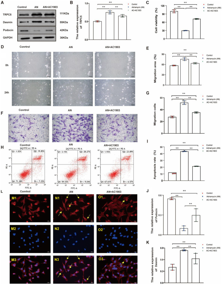
Figure 7.
TRPC5 inhibitor alleviates Adriamycin-induced podocyte injury. (A) Western blotting to assess the levels of TRPC5, Desmin and podocin protein in podocytes. (B) TRPC5 level was significantly reduced in the AN+AC1903 group compared with that in the Control and AN group (n = 3) (**p < 0.01). (C,D) Relative densities of Desmin and podocin protein expression normalized to GAPDH (n = 3) **p < 0.01. (E) Podocyte migration area increased in the AN+AC1903 (4.06 μM) group compared with that in the AN group, as analyzed using a scratch wound assay. Scale bar: 100 μm. (F) Quantitative analysis of the migration rates in (D) (n = 3) **p < 0.01. (G) Podocyte migration increased in the AN+AC1903 (4.06 μM) group compared with that in the AN group, as confirmed using the Transwell assay. Scale bar: 50 μm. (H) Quantitative analysis of the migrated cells in (F) (n = 3) **p < 0.01. (I) Apoptotic podocytes were determined using flow cytometry. (J) Relative apoptosis rate of podocytes following AN and AN+AC1903 (4.06 μM) treatments (n = 3) **p < 0.01. (K) Cell viability rate of podocytes in the AN+AC1903 (4.06 μM) group was higher than that in the AN group (n = 3) (**p < 0.01). (L) The yellow arrow indicates the lamellipodium (scale bar = 20 μM). M1/N1/O1 represents three groups of podocyte actin cytoskeleton; M2/N2/O2 DAPI represents DAPI staining of the three groups of podocyte nuclei. M3/N3/O3 represents the merge of M1/M2, N1/N2, and O1/O2.

