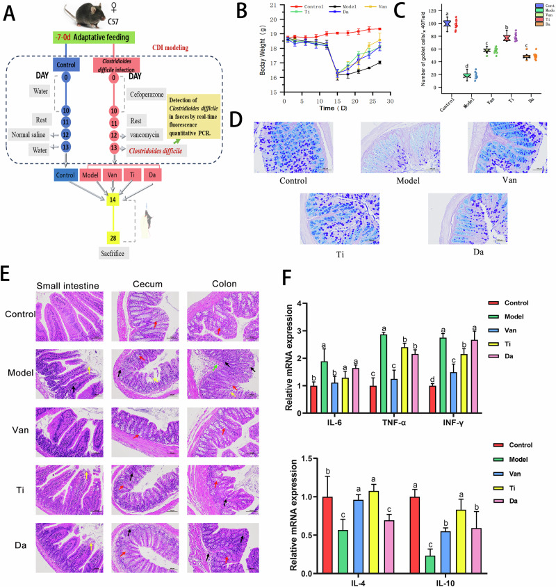
Fig. 1. Flowchart of animal experiment and Effects of peptides from E. japonicus and G. max on clinical symptoms and intestinal mucosal barrier in mice of CDI.
Different letters indicate significant difference between each group. (n = 5). A Flowchart of animal experiment. B Effects of peptides on the body weight in mice of CDI. C Counting the number of goblet cells in the colon of different groups of mice. D Effects of peptides on colonic Goblet cell in mice of CDI. (×200). E Effects of peptides on pathological changes of small intestine, cecum, and colon tissues in mice of CDI. (×200). Black arrow: inflammatory cell infiltration;Red arrow: goblet cells;Yellow arrow: mucosal epithelial cell erosion and detachment;Green arrow: submucosal edema. F Effects of peptides on colonic inflammatory factors. Pro-inflammatory factors: IL-6(Interleukin 6), TNF-α(Tumor Necrosis Factor-alpha), INF-γ:(Interferon -γ); Anti-inflammatory factors: IL-4(Interleukin 4), IL-10(Interleukin 10). Different lowercase letters represent significant differences between each other for the same indicator (P < 0.05), same as below.

