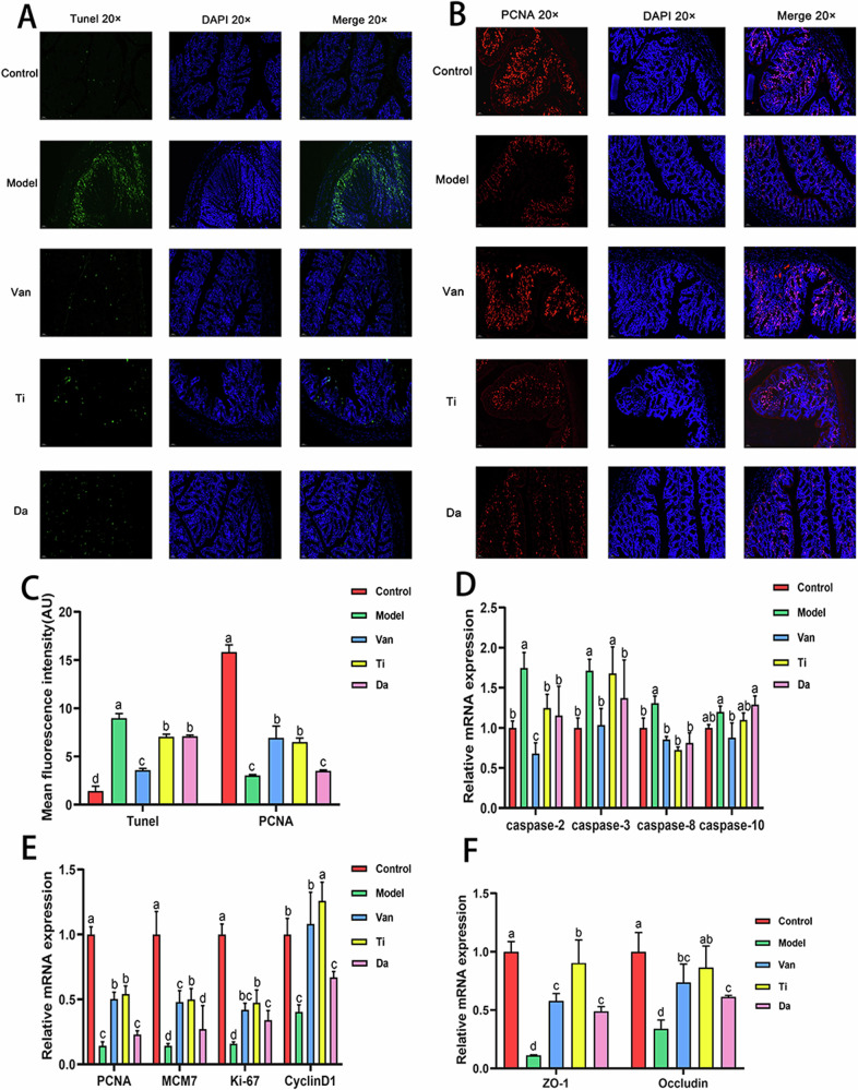
Fig. 3. Effects of peptides from E. japonicus and G. max on apoptosis and proliferation of colonic epithelial cells in mice of CDI.
Different letters indicate significant difference between each group (n = 5). A Colon immunofluorescence staining shows the presence and distribution of apoptotic cells and nuclei, magnification: (B) Existence and distribution of proliferating cells and nuclei, magnification. C Semi-quantitative analysis of fluorescence intensity of TUNEL and PCNA staining. D Effects of peptides on relative expression levels of apoptosis-associated genes in mouse colon epithelial cells. E Effects of peptides on relative expression levels of proliferation-associated genes in mouse colon epithelial cells. F Effects of peptides on the tight junctions of colonic epithelial cells.

