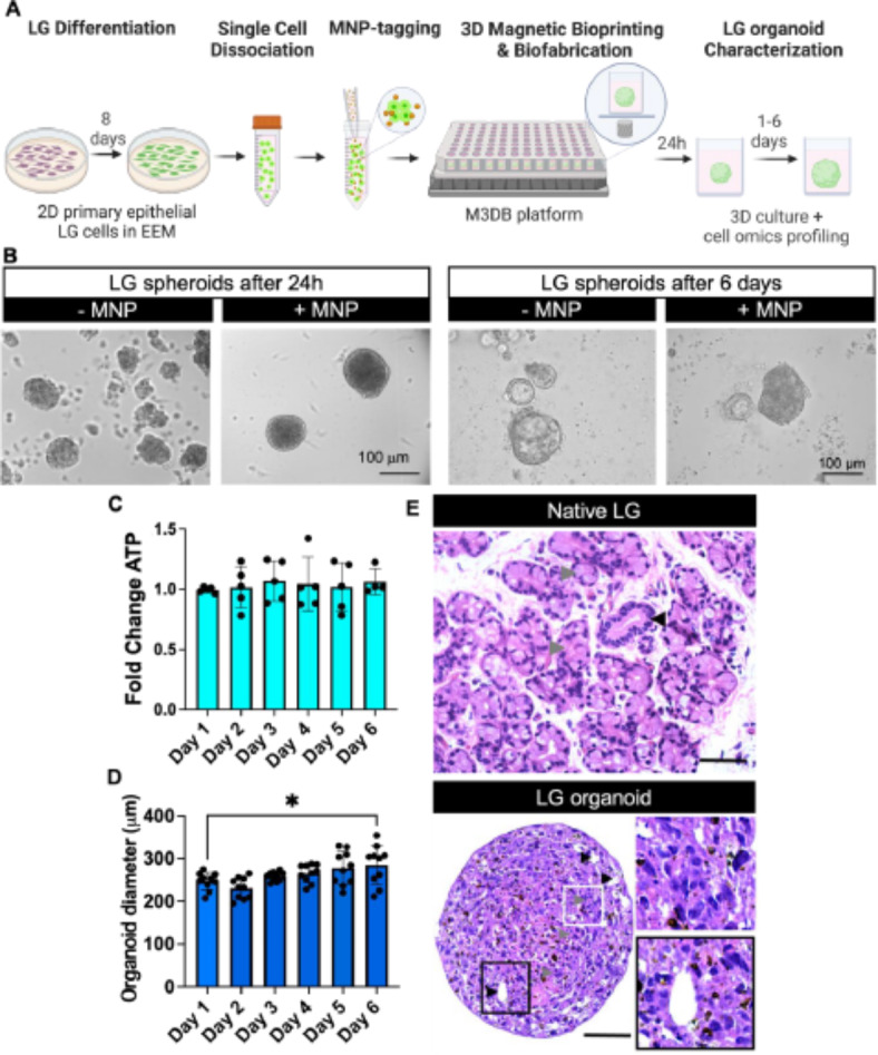
Fig. 1.

Epithelial morphology and viability of a M3DB-assembled LG organoid. (A) LG organoid biofabrication workflow steps. EEM epithelial-enriched media, MNP magnetic nanoparticles, M3DB magnetic 3D bioprinting/bioassembly. Created with BioRender.com. (B) Brightfield micrographs with phase contrast showing consistent spheroid formation in MNP-tagged LG primary cells. Scale bars: 100 μm. (C) Fold change in total ATP of each organoid was determined by a CellTiter-Glo 3D assay (n = 4–5) and all values were normalized to day 1 (baseline). The total ATP was deemed stable through 6 days of culture (n = 5). (D) After biofabrication, organoid diameter was determined and consistent up to day 3, after which there was wide variation in organoid size (n = 10). *p < 0.05 when compared to baseline or day 1, with one-way ANOVA with Dunnett’s post-hoc test. (E) H&E micrographs displayed a structural integrity and epithelial parenchyma with acini-like clusters (gray arrowheads, top white framed inset at higher magnification) and lumen-like regions (black arrowhead, bottom black framed inset at higher magnification) of the LG organoid at day 3 of culture resembling that of native LG fresh biopsy tissue. Inset is a focused area arising from black frame box. Scale bars: 50 μm.
