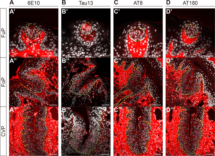
Fig. 1.
Detection of amyloid precursor protein (APP) and phosphorylated tau (p-tau) in taste buds. Representative confocal images of 6E10-immunoreactivity (IR) (A), Tau13-IR (B), AT8-IR (C), and AT180-IR (D) in fungiform (FuP) (A′~D′), folate (FoP) (A″~D″), and circumvallate papillae (CVP) (A‴~D‴) of 8-week-old C57BL/6 mice. Yellow dashed lines demarcate the epithelial boundaries. Immunofluorescent (IF) signals (red) and DAPI (white). Scale bars=50 μm.

