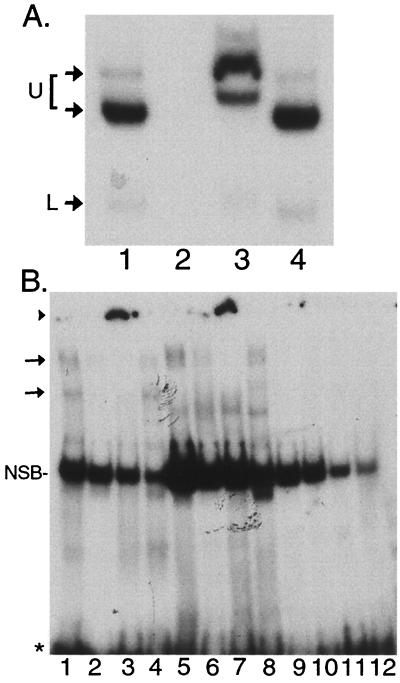
FIG. 7.
Supershift analyses of F-LANA1 and PEL cell nuclear extracts with TR-13. (A) Anti-FLAG antibody supershifts F-LANA1 TR-13 complexes. In vitro-translated F-LANA1 was incubated with TR-13 probe for all lanes. A 50-fold excess of unlabeled TR-13 oligonucleotide (lane 2), monoclonal anti-FLAG antibody (lane 3), or isotype-matched control antibody (lane 4) was added 15 min prior to the addition of 50,000 cpm of TR-13. Arrows indicate specific gel shifts. U, upper F-LANA1 shifted complex seen in Fig. 6B and C, which is resolved into two complexes here after a longer gel run; L, lower F-LANA1 gel-shifted complex. Data shown are representative of three experiments. Free probe was run off the gel. (B) LANA1 from PEL cells gel shifts TR-13. EMSA was performed using TR-13 probe and nuclear extracts from BCBL-1 (lanes 1 to 4), BC-1 (lanes 5 to 8) or (uninfected) BJAB cells (lanes 9 to 12). A 50-fold molar excess of unlabeled TR-13 (lanes 2, 6, and 10), anti-LANA1 monoclonal antibody (lanes 3, 7, and 11), or control antibody (lanes 4, 8, and 12) were included in incubations prior to the addition of 50,000 cpm of TR-13 probe. The arrows indicate specific gel shifts, and the arrowhead indicates supershifted probe near the gel origin (lanes 3 and 7). NSB, nonspecific band. Free probe is indicated by an asterisk. Data shown are representative of three experiments.

