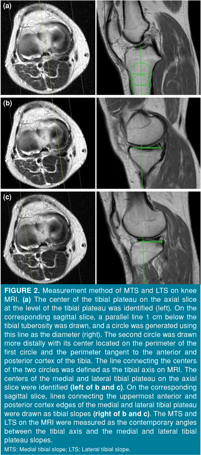
Figure 2. Measurement method of MTS and LTS on knee MRI. (a) The center of the tibial plateau on the axial slice at the level of the tibial plateau was identified (left). On the corresponding sagittal slice, a parallel line 1 cm below the tibial tuberosity was drawn, and a circle was generated using this line as the diameter (right). The second circle was drawn more distally with its center located on the perimeter of the first circle and the perimeter tangent to the anterior and posterior cortex of the tibia. The line connecting the centers of the two circles was defined as the tibial axis on MRI. The centers of the medial and lateral tibial plateau on the axial slice were identified (left of b and c). On the corresponding sagittal slice, lines connecting the uppermost anterior and posterior cortex edges of the medial and lateral tibial plateau were drawn as tibial slopes (right of b and c). The MTS and LTS on the MRI were measured as the contemporary angles between the tibial axis and the medial and lateral tibial plateau slopes.<br> MTS: Medial tibial slope; LTS: Lateral tibial slope.

