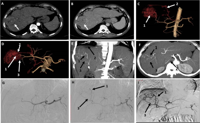
Fig. 5.
A patient with segment VII HCC measuring 6 × 5 cm. (A) and (B) Axial CT images in the arterial and portal phases, respectively, showing early arterial enhancement of the lesion with early contrast washout. (C) and (D) are 3D reconstructed angiography images in the AP view and cranio-caudal view, respectively, showing three possible feeding arteries numbered 1, 2, and 3. (E) and (F) Coronal and axial MIP CT images showing the three possible feeding arteries and the origin of artery no. 2 (right inferior phrenic artery) from the origin of the celiac trunk. (G) DSA of the celiac trunk using a 5fr catheter showing branches of the celiac trunk with no detectable contrast opacification of the right subphrenic artery (feeding artery no. 2). (H) and (I) DSA of the Celiac artery and CHA using a 5fr catheter showing the other two possible feeding arteries 1 and 3. Observer 1 detected arteries 1, 2 and 3 in 3D images, while observer 2 detected only arteries 1 and 3. Both observers identified arteries 1 and 3 in the DSA images. The three arteries are true feeding arteries for the lesion according to the ground truth

