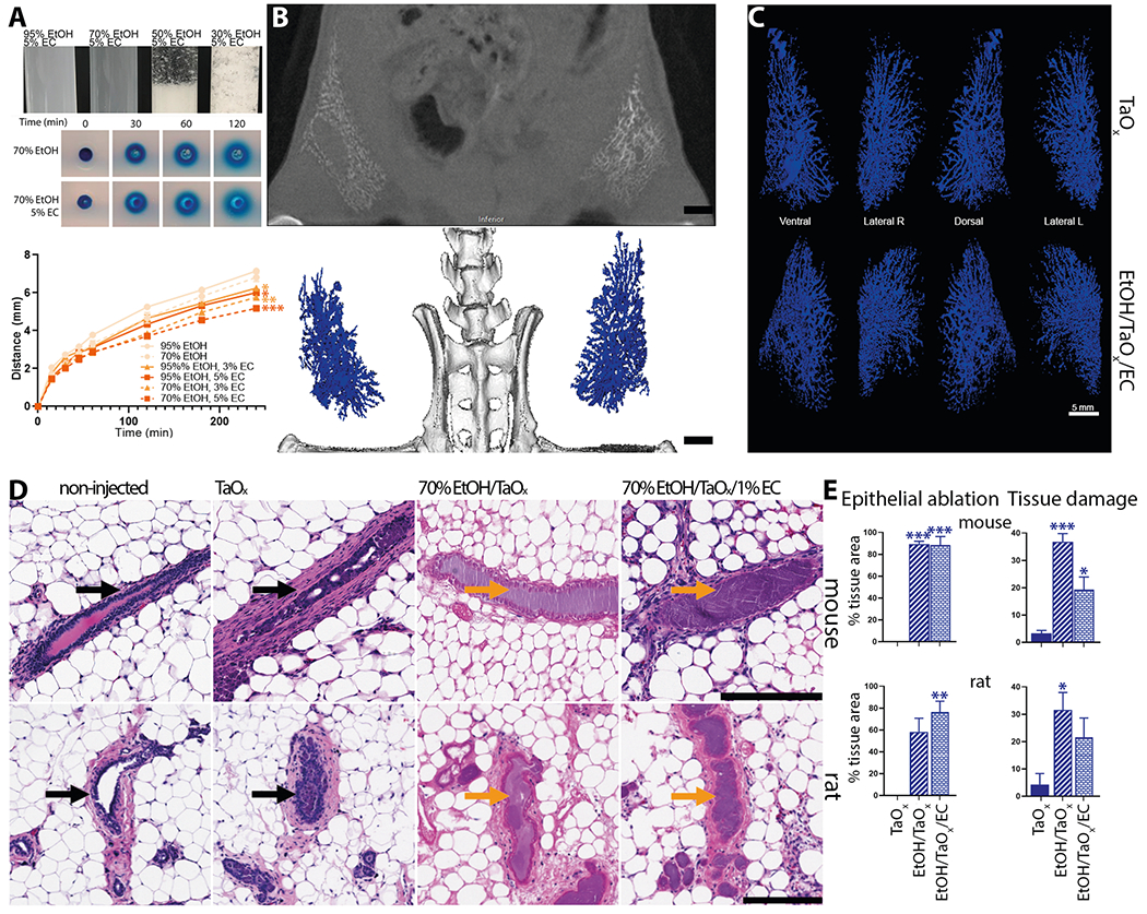
Fig. 7. Compatibility and scalability of TaOx-containing solutions.

A Indicated blue dye-containing solutions were dispensed into 1% agarose-casted 5-mm circular cylindrical channels (tissue phantoms). The distance the blue dye front traveled from the edge of each channel (x) was plotted over time (t). All solutions fit (R2 > 0.99) Fick’s equation x = (4Dt)½, where D is the diffusion constant. Asterisks indicate p value of unpaired Welch’s t-test of each solution compared to 95% EtOH (* < 0.01, ** <0.001, *** <0.0001). B, C Ductal trees of abdominal mammary glands were infused with 250 μl of indicated TaOx-containing solutions (18 mg Ta/ml). B Representative microCT slice of the lower body of an animal is shown immediately after last ID injection. Scale bar is 10 mm. C 3D reconstruction of manually segmented mammary gland per condition is shown at different views. 3D reconstruction was thresholded to include only voxels with a HU value of >300. Scale bar is 5 mm. D Representative H&E staining of mouse and rat mammary gland 3 days after ID injections of indicated TaOx-containing solutions (18 mg Ta/ml). Intact (black arrow) and ablated ducts (orange arrow) are indicated. Scale bar is 200 μm in images at different magnification. E Morphology-driven quantitation of epithelial ablation (anucleate cells, cytoplasmic hypochromia) and tissue damage, which includes fibrosis, inflammation and scarring resulting from ablative effects of 70% EtOH as well as immune cell-mediated foreign object reaction to clear nanoparticle-based contrast agents. Asterisks indicate p value of unpaired Welch’s t-test of each solution compared to TaOx solution.
