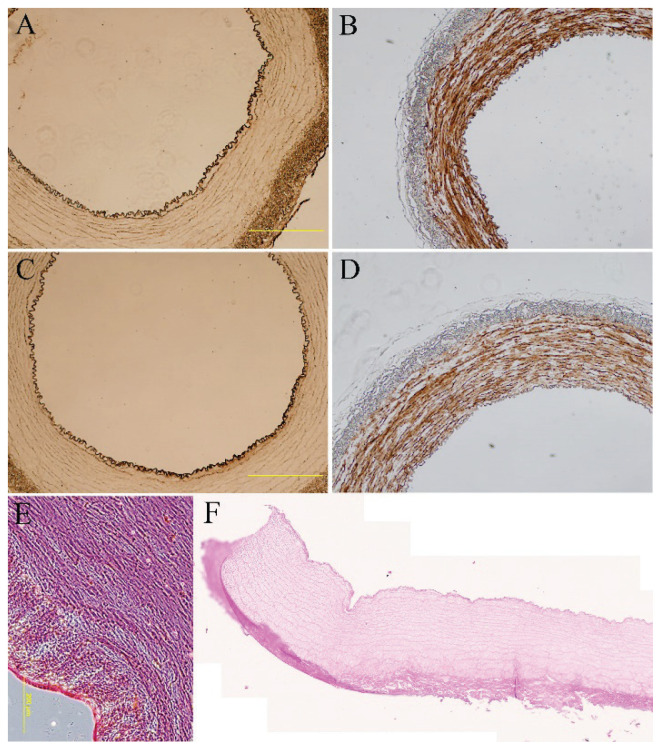
Fig. 2.
Immunohistological staining of von Willebrand factor (A, C) and calponin (B, D) of endothelialized porcine carotid artery that had been decellularized and seeded with human adipose tissue-derived stem cells (A, B) or human bone marrow-derived stem cells (C, D) for 14 days and human umbilical vein endothelial cells for two days of in vitro culture. Hematoxylin-eosin staining of control native (E) and decellularized arteries, tile scan (F). A–E: Olympus IX 71 epifluorescence microscope, DP80 digital camera; A–D: obj.×4, scale bar = 500 μm; E: obj.×10, scale bar = 200 μm; F: ZEISS Axio Scan. Z1 Slide Scanner, obj.×20.

