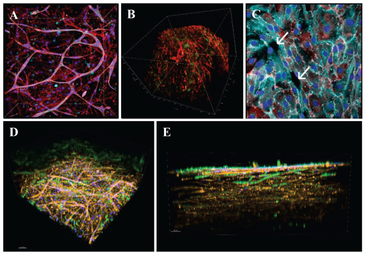
Fig. 4.
A: Capillary-like network formation in a collagen hydrogel with embedded HUVECs and ADSCs immigrating from an underlying fibrin-modified electrospun nanofibrous polylactide membrane after 14 days in culture. B: Immunofluorescence staining of β-actin (red) and calponin (green) in human ADSCs in collagen gels on day 14 of culture in EGM-2 medium. C: A monolayer of HUVECs originating from the openings (arrows) of capillary-like structures on the surface of the collagen hydrogel. A, C: Both cell types were stained with phalloidin for cytoskeletal F-actin (conjugated with Atto 488; all images; red), and with DAPI for cell nuclei (all images; blue). The CD31 membrane marker of HUVECs was visualized by immunofluorescence (Alexa 633; turquoise). D, E: bottom and side view of the construct, where the collagen hydrogel was enriched with fibronectin (10 μg/ml). Both HUVECs and ADSCs were stained with phalloidin for the cytoskeletal F-actin (red), and with DAPI for the cell nuclei (blue). Von Willebrand factor, a marker of HUVECs, was visualized by immunofluorescence (green). Scale bar 50 μm. A, C, D, E: Dragonfly 503 spinning disk confocal microscope, obj. ×20 (A, D, E), obj. ×40 (C). B: Zeiss Z.1 light-sheet microscope, obj. ×10 for excitation, obj. ×20 for detection, zoom ×0.4 tile scans.

